examination

Achilles Tendon Rupture

AnatomyAchilles tendon anatomy


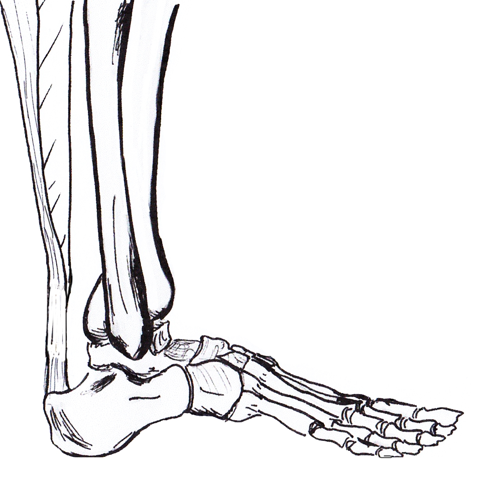
Gastrocnemius tendon 10-25 cm long

- soleus 3-10 cm

- inserts superior calcaneal tuberosity

- fibres spiral 90°

- fibres that lie medially in proximal portion become posterior distally

- allows elastic recoil & energy storage


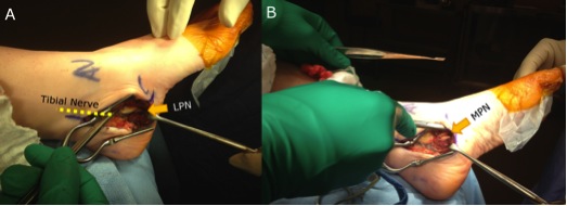
Plantaris present in 90% population

- medial to T Achilles


Poor blood supply midportion

- mesotenal vessels

Calcaneo Cavus

Definition


Pure Cavus Deformity characterised by 

- dorsiflexion of Calcaneus 

- plantarflexion of Forefoot


Aetiology


Weakness of Tendoachilles


Usually neuromuscular

- Polio (Most common worldwide) 

- Spina bifida

- CP (can be due to overcorrection of T Ach)

- Spinocerebellar Degen (Friedreich's Ataxia)

Tendonitis / Tendonopathy

Anatomy


Lateral compartment of leg

- run through retromalleolar groove

- pass superior and inferior to peroneal tubercle

- covered by inferior peroneal retinaculum


Peroneus longus

- origin lateral condyle of tibia and head fibula

- tendon PL superficial and inferior to brevis in retromalleolar groove

- runs in cuboid groove 

- insert plantar surface base of 1st MT and lateral aspect medial cuneiform

Dislocation

Definition


Anterior displacement of peroneal tendons out of peroneal groove


Epidemiology


Most common in young adults


Acute injury often missed 


Aetiology


Congenital


3 % neonates

- resolves spontaneously


Traumatic  


Occurs following sporting activities

Cervical Myelopathy

DefinitionCervical Myelopathy MRI


Spinal cord dysfunction
- extrinsic compression of the cord or its vascular supply
- caused by degenerative disease of spine


Epidemiology


Most common spinal cord dysfunction in patients > 55 years old
 

C5/6 commonest level