Management
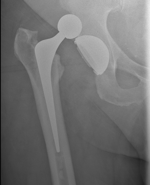
Timing
Early < 3 - 6 months
- most common
- excessive hip position by patient
- before adequate muscle control & soft tissue healing
Secondary 6 months - 5 years
- represents majority of recurrent dislocations
Early < 3 - 6 months
- most common
- excessive hip position by patient
- before adequate muscle control & soft tissue healing
Secondary 6 months - 5 years
- represents majority of recurrent dislocations
Benign intramedullary cartilage lesion
Movement of iliopsoas tendon over femoral head / iliofemoral ridge / iliofemoral ligament
Can be seen following THA with cup impingement
Audible or palpable snap in the groin
Hip moves
- from flexed / abducted / externally rotated position
- to extended / internally rotated position
10% of the population - usually painless
Athletes with increase activity / distance
Women with eating disorders / amenorrhea
Compression / inferior neck
- < 50% protective weight bear
- > 50% emergent ORIF
Tension side / superior neck
- emergent ORIF
1. Capsular avulsions
2. Body / Nutcracker fracture
Epidemiology
- rare
Mechanism
- forced eversion / abduction of forefoot
- cuboid crushed between 4th and 5th MT and calaneum
Pathology
- displaced cuboid fracture with subluxation of tarsus
Coronal plane fracture of distal femoral condyle
- intra-articular
- often only attachment is posterior capsule
Rare
Usually a severe valgus trauma
Insufficiency fracture
- secondary to exceeding fatigue threshold
- usually of second or third MT shaft
Onset of new and very intense / strenuous physical activity
- i.e. new army recruits / dancers
Women with postmenopausal osteoporosis
Cavus feet
Children < 6
- entire distal humerus physis is displaced
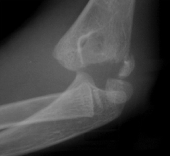
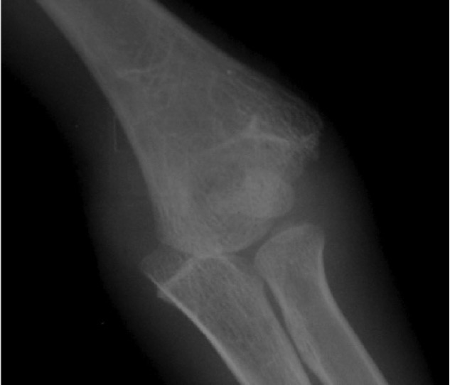
Distal physis not ossified < 1 year
- may be a difficult diagnosis
> 10 mg / dl
- must be corrected for albumin
Malignancy
- multiple myeloma / lung cancer / breast cancer
Hyperparathyroidism
- elevated PTH
High mortality associated with hypercalcaemia of malignancy
40% albumin bound
50% ionised and active
Fall in level promotes tetanus
Chvostek sign
- tapping masseter muscle induces spasm
Trousseau Sign
- flexion of thumb & wrist with extension of fingers
Carpopedal Spasm
Prolonged QT interval on ECG
1. Vit D Deficiency