Definitions
Patellofemoral Pain Syndrome
- anterior knee pain in the absence of cartilage damage
- adolescents
- female > male
- maltracking
- patella tilt / Lateral patella pressure syndrome
Chondromalacia patella
- damage to the articular cartilage of the patella / trochlea causing anterior knee pain
- trauma
- acute PFJ dislocation
- chronic maltracking
- patella tilt / Lateral patella pressure syndrome
Epidemiology
- systematic review of anterior knee pain
- incidence general population 23%
- incidence adolescents 29%
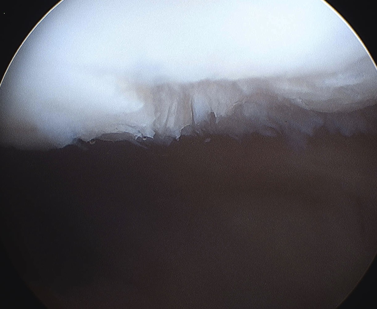
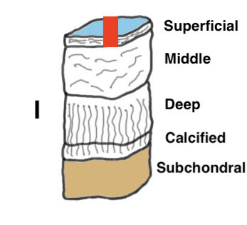
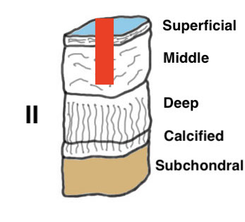
International Cartilage Research Society Arthroscopy (ICRS) Classification
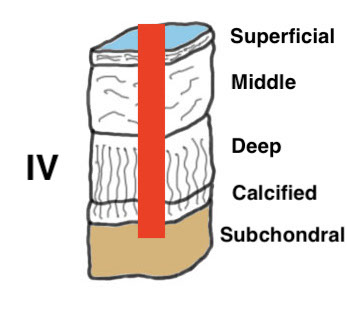
Grade I: Nearly normal - soft indentation / superficial fissures and cracks
Grade II: Abnormal - cartilage lesions < 50% of cartilage depth
Grade III: Severely abnormal - cartilage lesions > 50% of cartilage depth
Grade IV: Severely abnormal - cartilage lesion down to subchondral bone
Grade I: Softening
Grade II: < 50% of cartilage depth
Grade III: > 50% cartilage depth
Grade IV: subchondral bone
Symptoms
Anterior Knee Pain - often worse with prolong sitting
Swelling
Signs
PFJ crepitus
Maltracking
Tight lateral retinaculum
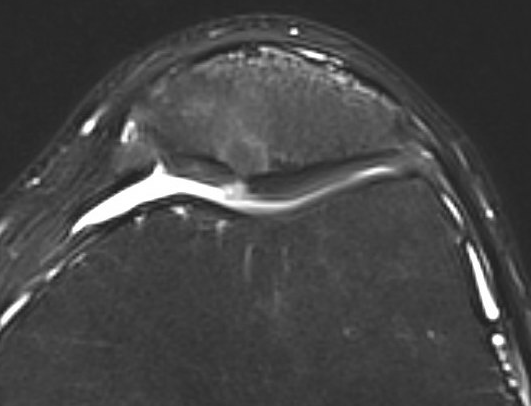
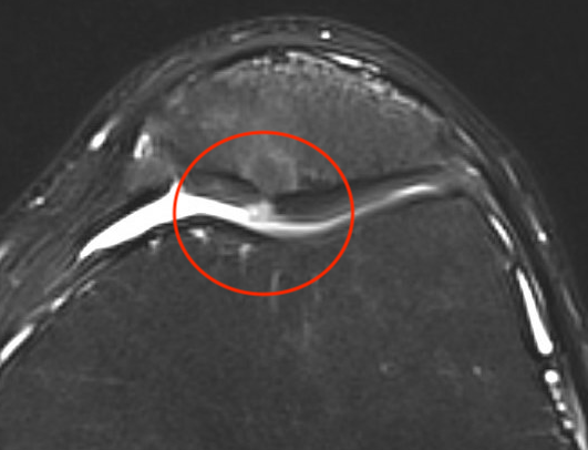
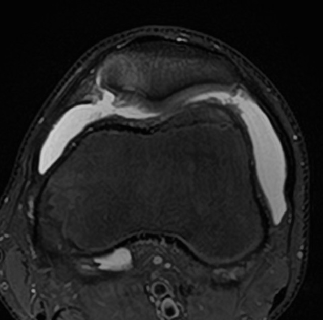
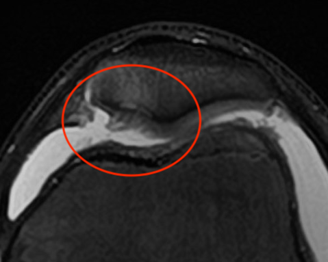
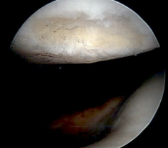
MRI
Non-operative Management
Physiotherapy
Quadriceps exercises
van der Heijden et al Cochrane Database 2015
- evidence for exercise therapy in reducing anterior knee pain
Patellofemoral knee braces
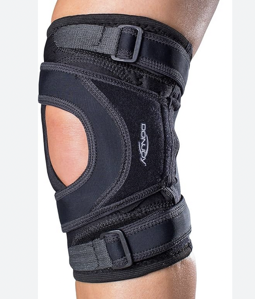
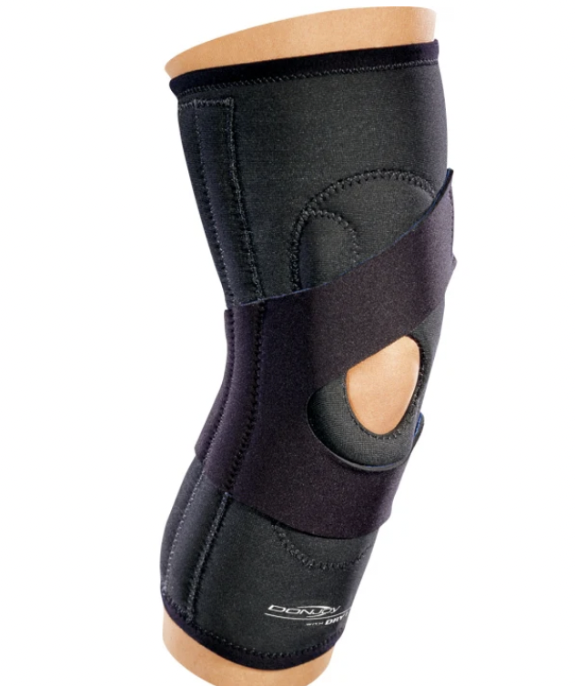
Culvenor et al Br J Sports Med 2020
- systematic review
- some evidence that braces can reduce AKP
Petersen et al Arch Orthop Trauma Surg 2016
- RCT of brace + exercise program versus exercise program alone
- 156 patients with patellofemoral pain syndrome
- better results with medially directed patella realignment brace
Taping
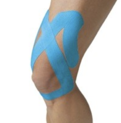
- systematic review of kinesio taping for patellofemoral pain syndrome
- evidence for improved pain, but not improved function
Neuromuscular stimulation
- RCT of 130 members of the military with PFPS
- use of electrical stimulation improved strength compared to strength training along
Injections
Hyaluronic acid
Hart et al Orthop J Sports Med 2019
- RCT of 86 patients with patellofemoral pain syndrome
- no efficacy of HA versus sham injections at 6 months
PRP
- meta-analysis of RCTs of injections for knee OA
- 10 RCTs and 1000 patients
- PRP superior to HA and saline at 12 months
- systematic review of PRP versus HA for knee osteoarthritis
- improved outcomes with PRP versus HA
Operative options for Patellofemoral pain syndrome
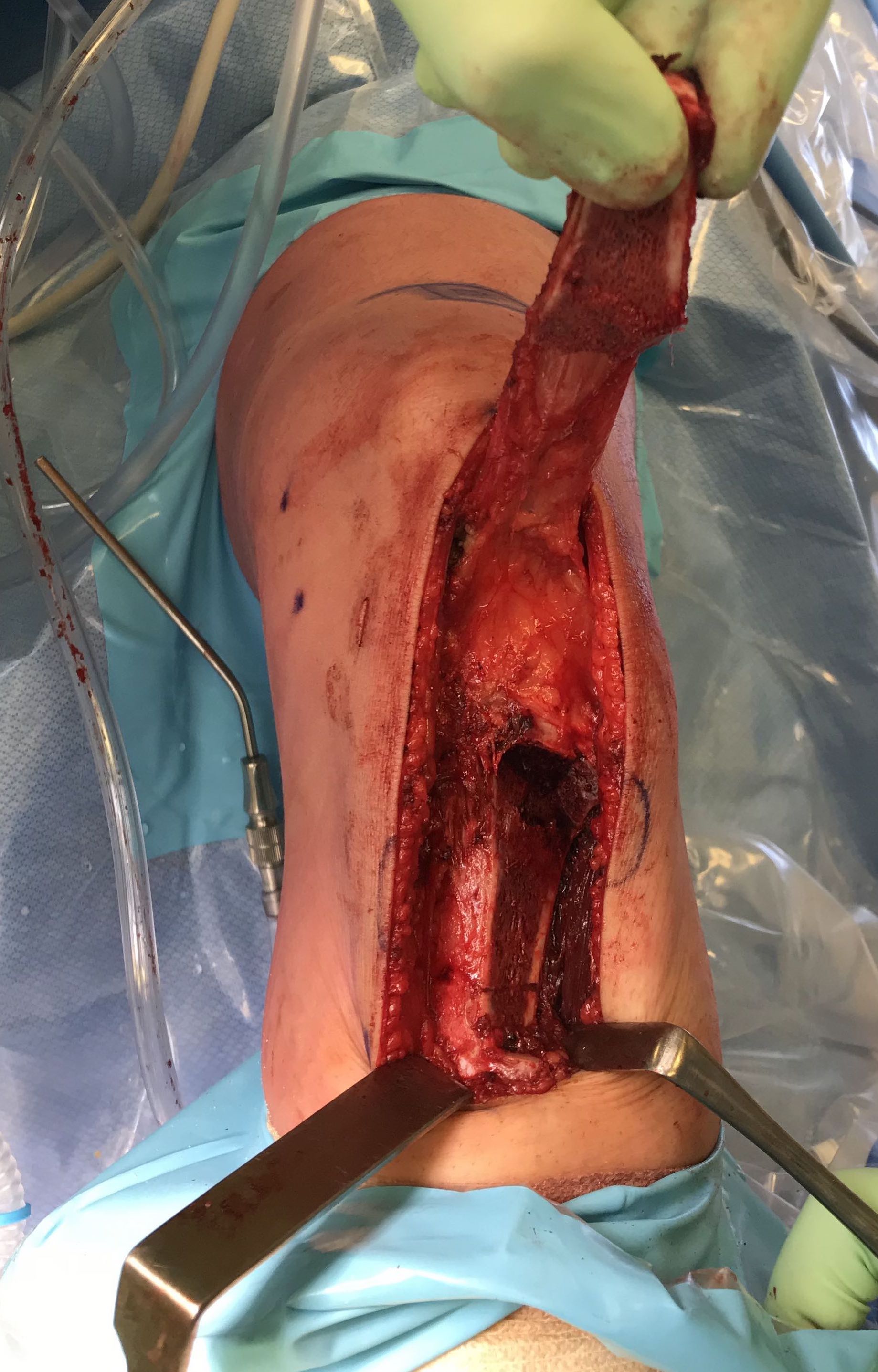
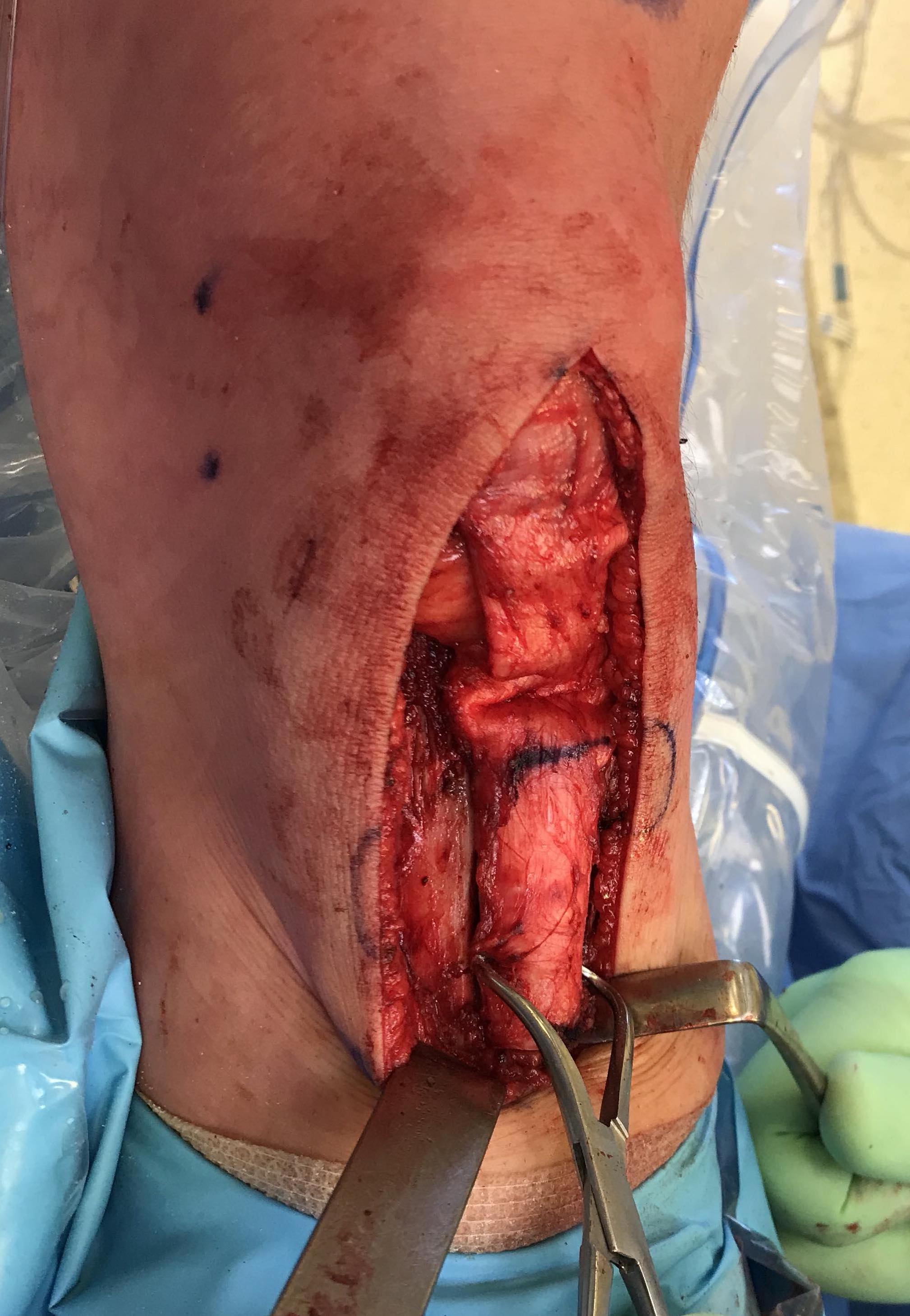
Tibial tuberosity Osteotomy
www.boneschool.com/tibialtuberosityosteotomy
Options
Medialization / Elmslie-Trillat
Anteromedialization / Fulkerson
Elevation / Maquet

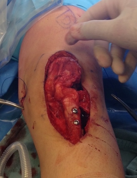
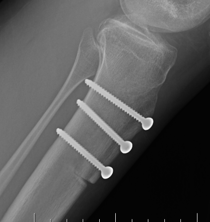
Fulkerson Maquet
Jack et al Bone Joint Res 2012
- 50 knees with normal tracking and CMP
- treated with Fulkerson / anteromedializaiton
- 72% good or excellent results
- 77 military patients undergoing a TTO for chondromalacia patella
- 63% returned to military duties
- 69 patients with grade I / II patellofemoral degeneration treated with anteromedialization
- 25% would not undergo the surgery again
Operative options for focal chondral defects of the patella
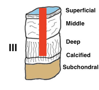
Arthroscopy
Debridement
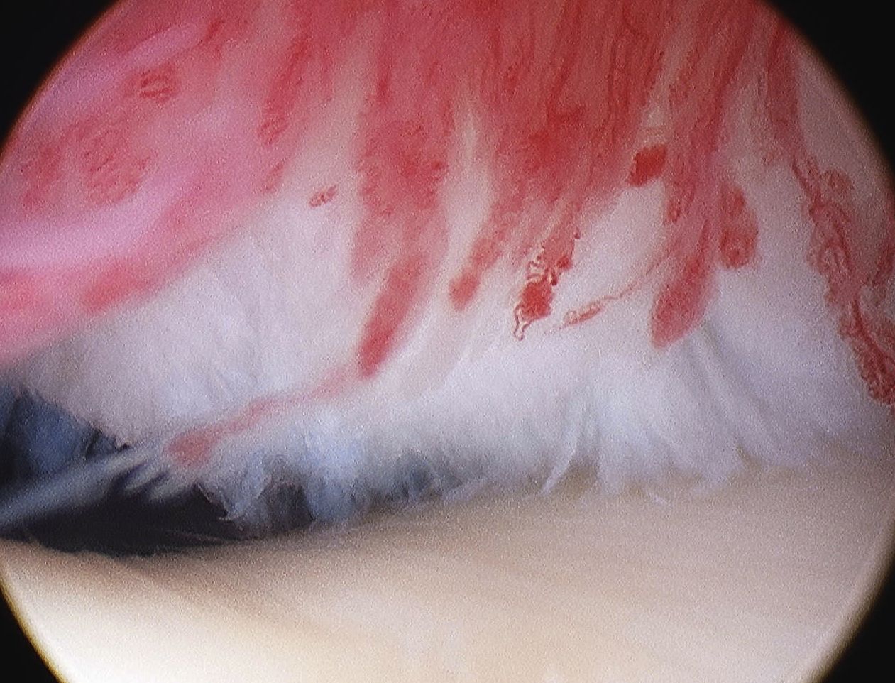
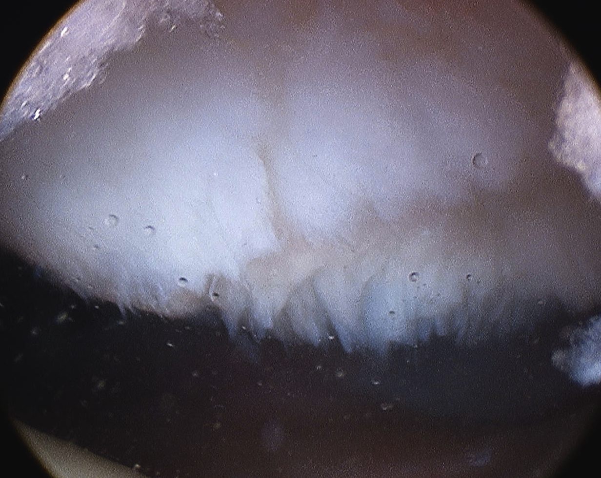
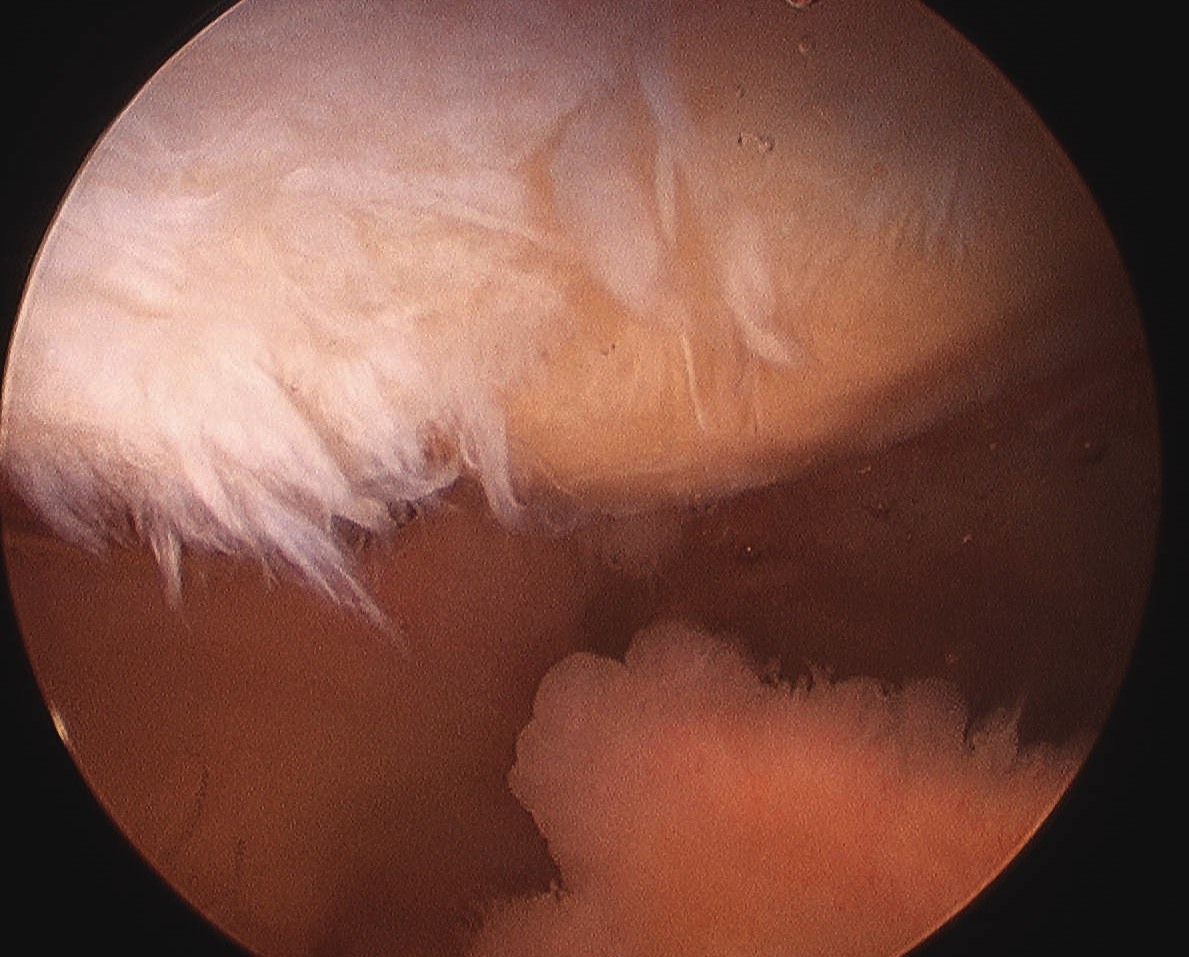
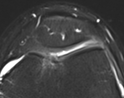
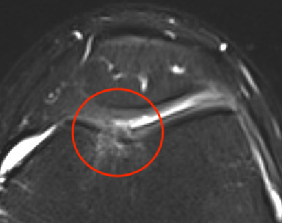
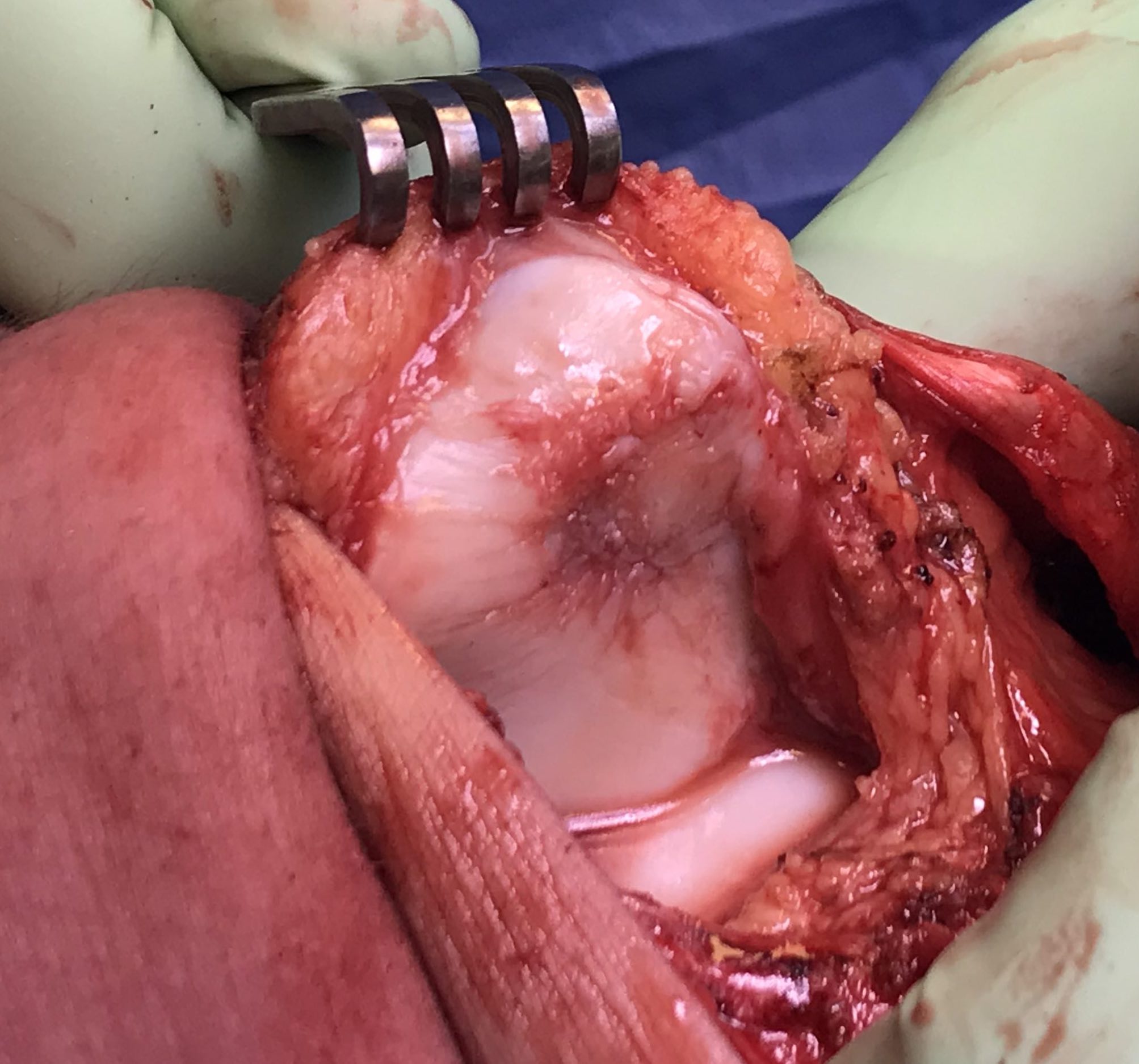
Debridement patella chondral flap
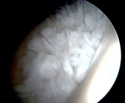
Debridement trochlea chondral damage
Federico et al Am J Sports Med 1997
- arthroscopic shaving in 36 patients with grade 2 or worse
- no malalignment
- only 50% good or excellent result
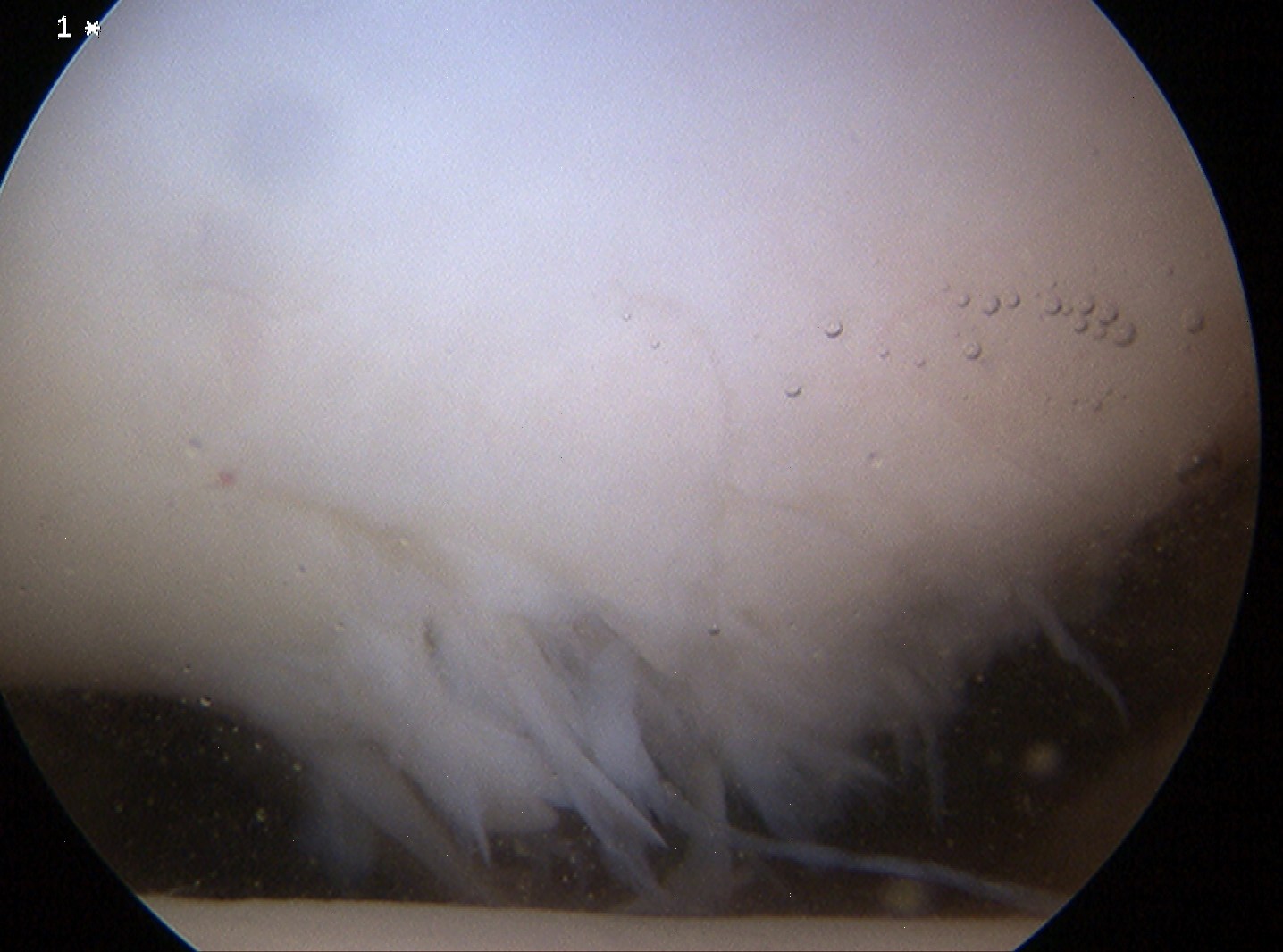
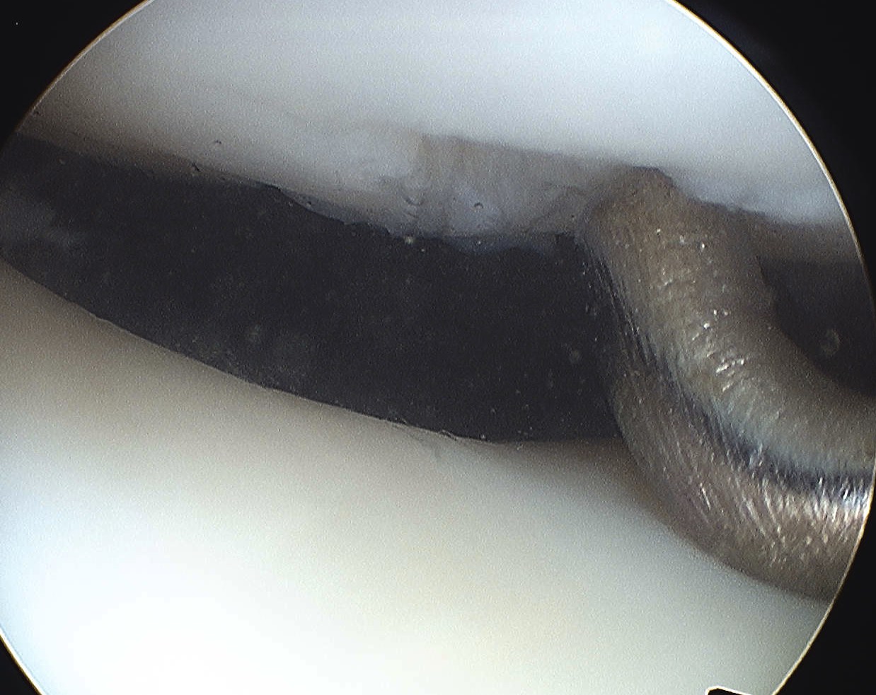
Microfracture

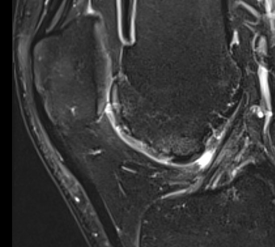
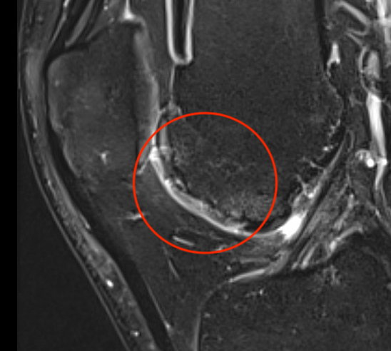
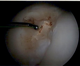
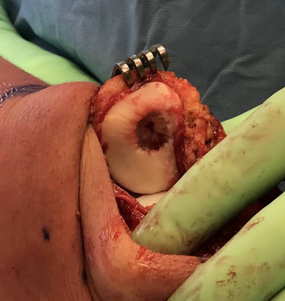
Microfracture of trochlea chondral defect
van Tuijn et al Cartilage 2023
- systematic review of microfracture
- worse outcomes in PFJ than in knee condyles
Cartilage restoration procedures
www.boneschool.com/chondraldefects
PFJ cartilage restoration procedures PFJ
- systematic review of PFJ cartilage lesions in 2000 patients
- clinical patient improvement of various restoration procedures
- MACI 83%
- OAT 78%
- OCA 71%
- AMIC 64%
- OCA had highest failure rate
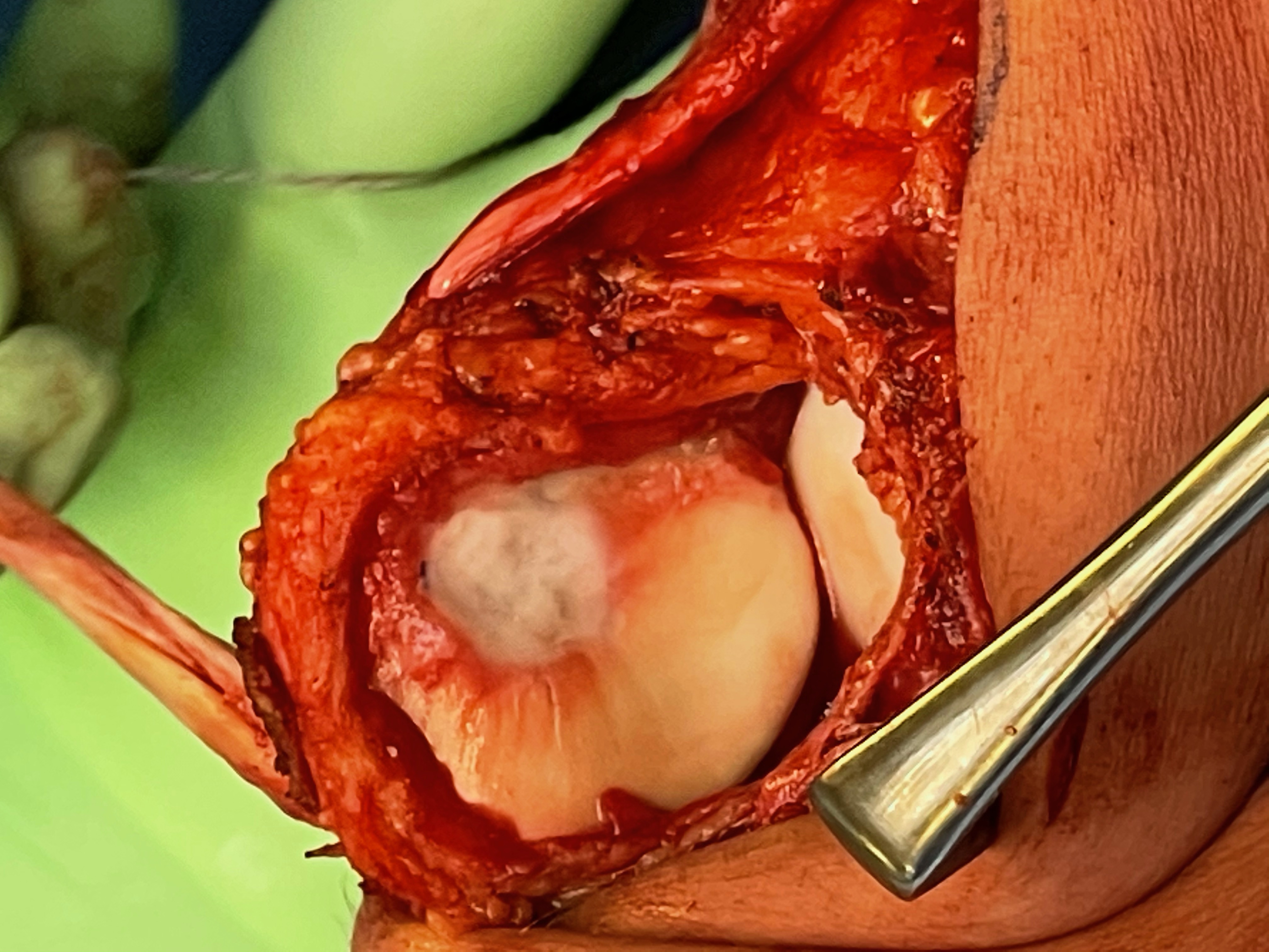
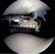
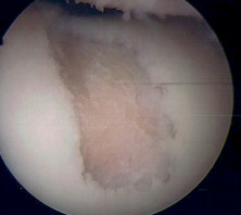
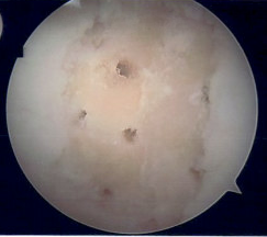
AMIC procedure of a full thickness patella lesion
Tibial tuberosity osteotomy (TTO)
- systematic review of patients with PFJ chondral lesions
- compare isolated ACI versus combined ACI and TTO
- better outcomes with ACI / TTO