
Diabetic Foot Complications
Diabetic foot infections
Diabetic foot ulcers
Charcot arthropathy
Epidemiology
McDermott et al Diabetes Care 2023
- incidence of diabetic foot ulcer 19 - 34%
- amputation risk of diabetic foot ulcer 20%
- 5 year mortality associated with diabetic foot ulcer 50 - 70%
Pathophysiology
1. Neuropathy
2. Peripheral vascular disease
Neuropathy
Most important factor in foot disease caused by
- glycosylation of nerves
- ischaemia
| Sensory | Autonomic | Motor |
|---|---|---|
| Stocking distribution |
20 – 40% of Diabetics |
Loss of intrinsic muscle balance |
|
Semmes Weinstein 5.07 monofilament - applies 10gm of force - tip pressed against skin until starts to bend - patient asked if they can feel it - 90% of patients who are able to feel won’t ulcerate |
Skin dry / scaly / cracked Easier access for bacteria |
Claw and hammer toes Increased risk of plantar ulcers |
Peripheral vascular disease
50% of diabetic foot ulcers have peripheral vascular disease
| Large Vessel Disease | Small vessel disease |
|---|---|
|
Different to non diabetic population - younger age - at or below knee - diffuse and longer occlusions |
Microangiopathy
|
|
Vascular claudication Hindfoot ulcers |
Delayed ulcer healing |
Management
Multidisciplinary teams
Musuuza et al J Vasc Surg 2020
- systematic review of multidisciplinary teams in management diabetic foot ulcer
- 94% studies demonstrate reduction in major amputations
Endocrinologist +/- diabetic nurse - glycemic control crucial
Podiatrist - non-surgical debridement / orthoses
Plaster technicians - total contact cast
Vascular surgeon
Orthopedic surgeon - total contact cast / surgical debridement / foot reconstruction / amputation
Infectious disease consultant - infections / non healing ulcers
Diabetic Foot Care
Daily foot hygiene
No walking barefoot
Immediate attention to blisters / ulcers
Custom shoes / orthoses
Diabetic foot ulcers
Wagner Classification
| Grade 0 | Grade I | Grade II |
|---|---|---|
| Pressure area | Superficial ulceration |
Deep ulceration Probes to tendon / capsule |
| Footwear modification |
Local treatment Footwear modification |
Total contact cast |
 |
 |
 |
| Grade III | Grade IV | Grade V |
|---|---|---|
|
Deep ulceration + Secondary infection |
Partial foot gangrene | Whole foot gangrene |
|
Amputation Hyperbaric oxygen |
Amputation | |
 |
 |
 |
University of Texas Classification
| Grade | Stage |
|---|---|
|
1 Preulcerative 2 Superficial Wound 3 Deep wound penetrating to capsule or tendon 4 Deep penetrating to bone or joint |
A Clean B Non ischemic Infected C Ischemic Noninfected D Ischemic Infected |
Perfusion
| Ankle Brachial Index (ABI) | Transcutaneous O2 Measurement (TcPO2) | Toe Blood Pressure | Angiogram |
|---|---|---|---|
|
ABI: Ankle / Brachial Systolic BP at ankle and arm Normal 0.9 - 1.3 |
Electrode placed on warmed foot Affected by edema/ infection / neuropathy |
Plethysmography | |
|
<0.9 suggests PVD May be falsely elevated by calcified vessels |
<25 mmHg = unlikely to heal | >30 mmHg = good wound healing potential |
- systematic review
- transcutaneous oxygen measurement predicts wound healing and amputation
- ABI predictive of amputation but not wound healing
Osteomyelitis
Diagnosis
- probe to bone
- ESR > 70
- Xray signs of destruction
- MRI


Forefoot - diabetic foot ulcer with evidence of underlying osteomyelitis
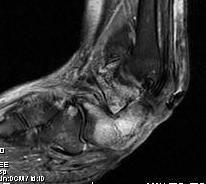
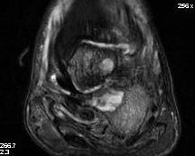
Hindfoot - calcaneal osteotomyelitis
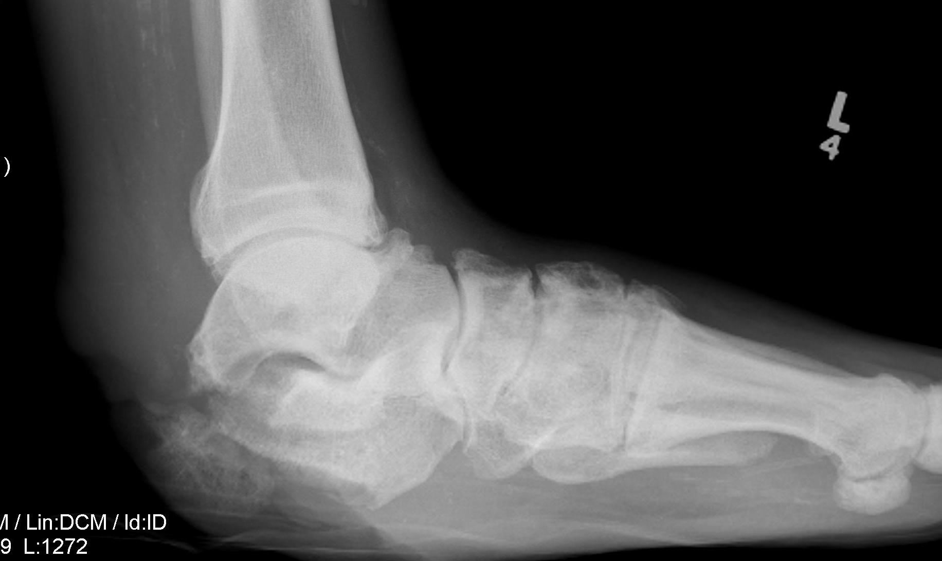
Charcot arthropathy


Midfoot ulcer with evidence of underlying Charcot arthropathy and midfoot collapse
www.boneschool.com/charcot-foot
Prognosis
- 154 diabetic foot ulcers
- 73% < 1 cm2
- ulcers healing at 12, 20, and 52 weeks were 59%, 71%, and 87%,
- 299 infected diabetic foot ulcers with 1 year follow up
- 15% 1 year mortality
- healing rate 46% with 10% recurrence
- amputation rate 17%
Nonoperative management
Options
Treat infection
Wound care / ulcer debridement
Offload foot - orthotics / total contact casts / CROW walkers
Infection
Pathogens
Widatalla et al Diabet Foot Ankle 2012
- 1800 diabetic foot infections and 330 diabetic foot osteomyelitis
- Staphylococcus aureus (33.3%), Pseudomonas aeruginosa (32.2%), Escherichia coli (22.2%)
Wound care and Ulcer debridement
Wound care
Modern dressings that absorb exudate and keep environment moist
- hydrogels / alginate
- silver dressings - antibacterial
Debridement
Wilcox et al JAMA Dermatol 2013
- 150,000 wound debridements
- increased healing with weekly (55%) versus less frequent debridement (28%)
Negative pressure therapy
Blume et al Diabetes Care 2008
- RCT of 342 patients with diabetic foot ulcers
- moist wound care +/- negative pressure therapy
- increased wound closure with negative pressure therapy (43% v 29%)
Hyperbaric oxygen
Oley et al Plastic Reconstr Surg Global Open 2024
- systematic review of hyperbaric oxygen for diabetic foot ulcers
- hyperbaric ulcers improved healing and prevent amputation
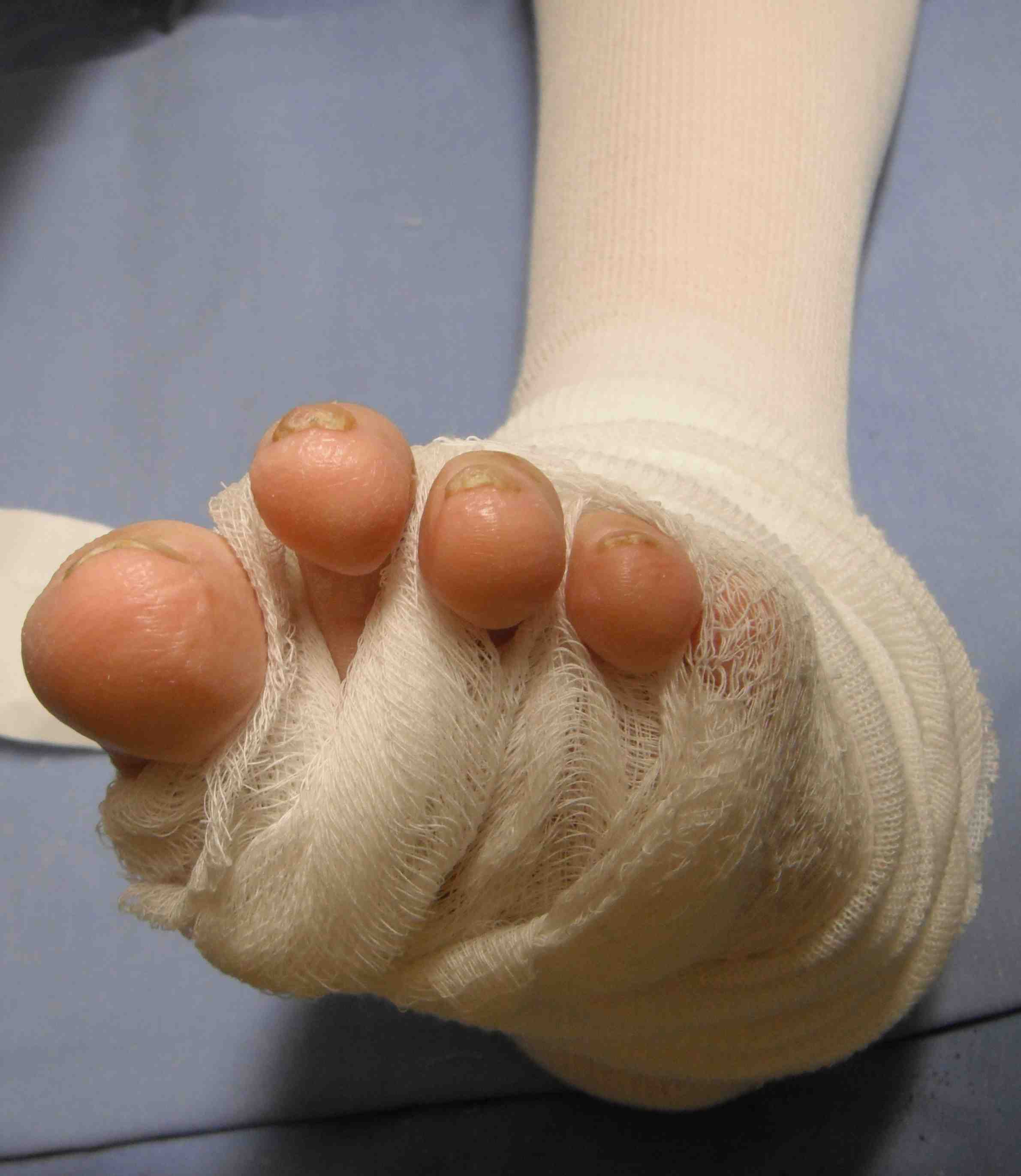
Offload ulcers
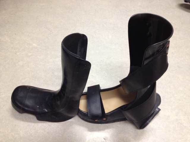
Total contact cast / Non removable walkers / removable walkers
- spread out force over a larger area
- can reduce pressure by as much as 80 - 90%
Indications
- superficial ulcers
- midfoot / forefoot ulcers (TCC don't reduce heel pressure)
- post surgery


Total contact cast
Results
Lazzarini et al Diabetes Metabol Res 2024
- systematic review of 194 studies
- increased wound healing with non removable devices (TCC) 82% versus removable 66%
- likely due to compliance issues
Nabuurs et al Diabetes Care 2005
- Total contact cast and ulcer healing in 98 patients
- neuropathic ulcers 90%
- infected neuropathic ulcers 87%
- neuropathic ulcers with peripheral artery disease 69%
- neuropathic ulcers with peripheral artery disease + infection 36%
Operative management
Options
Revascularization
Surgical debridement for osteomyelitis
Soft tissue releases - tendoachilles lengthening, toe flexor tenotomy
Bony realignment www.boneschool.com/charcot-foot
Amputations www.boneschool.com/diabetic-amputations
Surgical debridement of osteomyelitis
Indications
Osteomyelitis with failure of wound care / antibiotic therapy
Location
Osteomyelitis most common in forefoot, amputation most common with hindfoot osteomyelitis
Faglia et al Foot Ankle Int 2013
- 350 diabetic foot ulcers with osteomyelitis
- forefoot 86%, 8% midfoot, 7% hindfoot
- transtibial amputation 0.33% forefoot, 19% midfoot, 52% hindfoot OM
Surgery versus antiobiotics
Lazaro-Martinez et al Diabetes Care 2014
- 37 patients with diabetic foot ulcer and osteomyelitis
- RCT of antibiotics versus surgery + antibiotics in 37
- 75% primary wound healing + 17% minor amputations with antibiotics alone
- 86% wound healing + 14% minor amputations with surgery + antibiotics
Fractures in Neuropathic / Diabetic feet
Principles
1. Augment ankle ORIF
2. Double time for sutures
3. Double immobilization period
4. Brace for 1 year after surgery to prevent Charcot arthropathy