Femoral Artery
Enters thigh
- midway between ASIS and pubic symphysis
- in femoral triangle
- covered only by skin and fascia
- NAVY (nerve / artery / vein / Y fronts)
- in femoral sheath with femoral vein (transversalis fascia and psoas fascia)
- femoral nerve outside sheath / under the iliac fascia / lateral
Femoral triangle
Anatomy
- inguinal ligament superiorly
- sartorius laterally
- adductor longus medially
- floor is iliopsoas, pectineus and adductor longus
Femoral vein
- enters thigh posterior to femoral artery
- comes to lie medially
- receives great saphenous vein anteromedially
- in femoral triangle / just below femoral sheath
Femoral artery
- 4 branches under inguinal ligament
- superficial circumflex iliac
- superficial epigastric
- superficial and deep external pudenal
Enters adductor canal / subsartorial canal
Anatomy
- sartorius is roof
- floor is longus then magnus
- contents artery, vein and saphenous nerve
- vein comes to lie posterolateral
- artery always between vein and nerve
Saphenous nerve
- exits between sartorius and gracilis
- pierces fascia
- runs with great saphenous vein
Surgical approach
- medial thigh incision
- reflect sartorius medially
- divide fascial roof
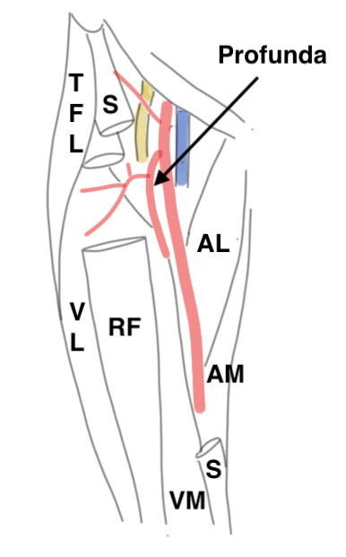
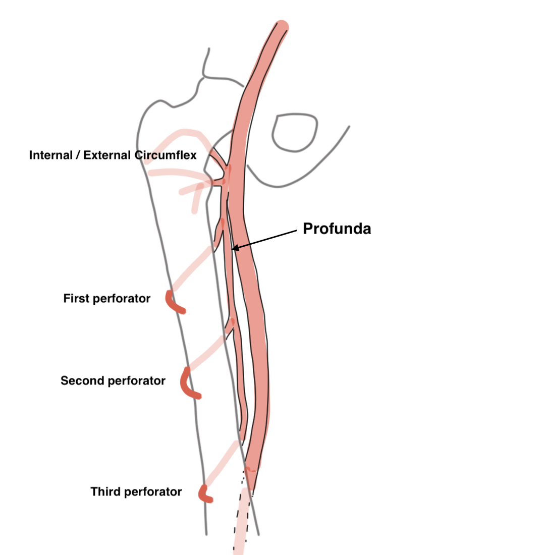
Profunda femoris
Artery to thigh muscles
- exits laterally at termination of femoral sheath
- 3-4 cm distal to inguinal ligament
- arises from lateral side
- passes between pectineus and adductor longus
- runs behind adductor longus
- runs down on adductor brevis and magnus
- gives 4 perforating branches
Perforators
- pass backwards through adductor magnus
- one above, second through, third and fourth below adductor brevis
Lateral circumflex
- lateral side of profunda
- passes laterally between branches of femoral nerve
- under sartorius and rectus
A. Ascending branch
- runs up between sartorius and TFL
- on vastus lateralis
- must be ligated in Smith Petersen approach
- supplies trochanteric anastomosis
- ends ASIS
B. Transverse branch
- passes across v. lateralis to wrap around proximal femur and supply cruciate anastomosis
C. Descending branch
- descends in groove between v. lateralis and intermedius
- travels with nerve to v. intermedius
Medial circumflex
Arises
- medial side profunda
- passes backwards between pectineus and psoas
- runs to back of femoral neck
Deep branch
- runs along inferior border of obturator externus
- emerges between obturator externus and quadratus femoris
- then runs over tendons of conjoint and piriformis
- supplys femoral head via posterior and superior femoral neck branches
Other branches
- anterior / posterior / transverse and trochanteric
Popliteal fossa
Boundaries
Superior: biceps laterally with semitendinosis / semimembranosus medially
Inferior: medial and lateral gastrocnemius
Floor: knee joint and capsule, popliteus
Popliteal artery
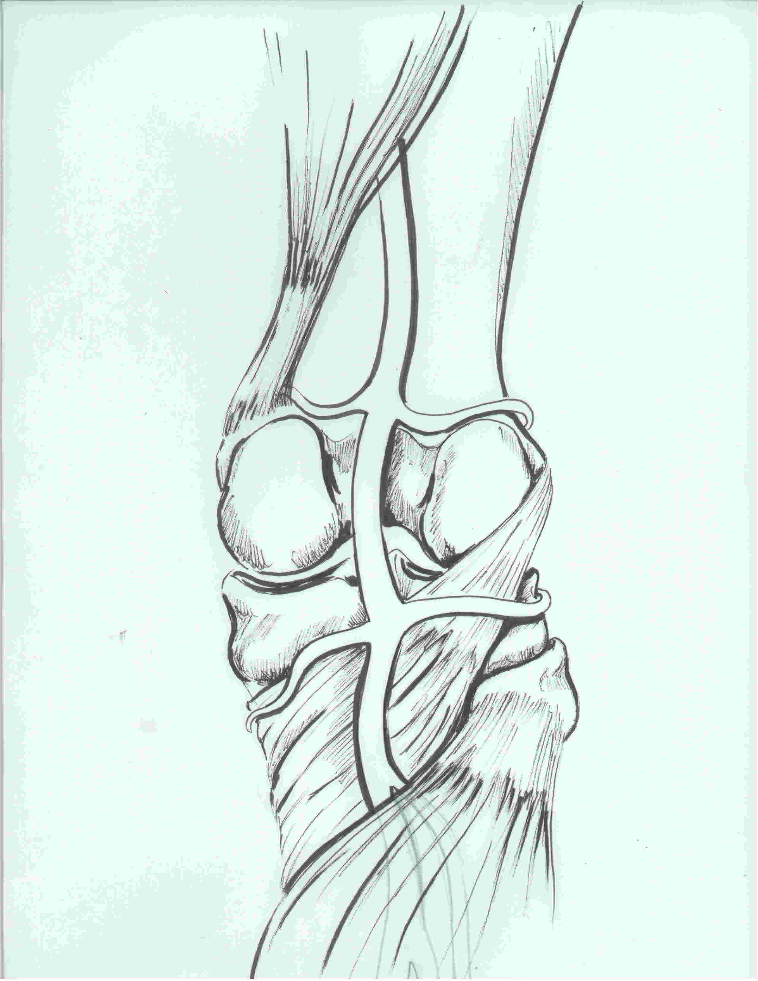
Anatomy
- deepest structure throughout
- enters deep and medial
- medial to femur via adductor hiatus
- bows laterally
- exits posterolateral to tibial nerve under fibrous arch of soleus
- returns to medial side
Popliteal vein
- always between artery and nerve
Divisions
- lower border of popliteus
- usually into anterior and posterior tibial artery (gives peroneal as a branch)
- sometimes into anterior and peroneal artery (
- sometimes into anterior / posterior and peroneal artery
Posterior Tibial Artery
Relations leg
- runs down on tibialis posterior behind deep intermuscular septum
- travels with tibial nerve
- at times between FHL and FDL
- gastronemius and soleus superficial
- gives peroneal artery as a branch
Relations ankle
- runs back of tibia and ankle joint to tarsal tunnel
- runs in groove behind medial malleolus between T posterior and FDL with tibial nerve
- passes deep to abductor hallucis and divides into medial and lateral plantar arteries
- found between first and second layer of the foot
- lateral plantar forms plantar arch by uniting with deep branch of the dorsalis pedis
Peroneal Artery
Origin
- usually branch of posterior tibial
- 2.5 cm below popliteus
Relations
- runs along medial aspect fibula
- between FHL and T posterior
Anterior Tibial Artery
Origin
- lower border of popliteus
Relations leg
- passes between two heads T posterior
- passes through aperature of interosseous membrane
- descends on interosseous membrane
- initially between T anterior and EDL
- eventually is crossed by EHL and crosses ankle between EHL and EDL
- runs with the deep peroneal nerve
- becomes dorsalis pedis under extensor retinaculum
- runs to interval between 1st and second MT