Anatomy
Unusual anatomic convergence of ilium, pubis and ischium
- covered entirely by hyaline cartilage
- except at acetabular fossa, which is the site of attachment of the ligamentum teres
- deepened by peripheral fibrocartilage labrum
2 column theory (Letournel and Judet)
Anterior Column
- superior pubic ramus
- anterior acetabular wall, anterior dome
- anterior iliac spines and anterior ilium
Posterior Column
- ischium
- posterior acetabular wall, posterior dome
- posterior ilium
Quadrangular Plate
Mechanism
Axial load applied through femur
- type of fracture depends on position of femur at time of injury
- IR - posterior column
- ER - anterior column
Examination
Resuscitation EMST
Detailed neurological exam
- sciatic nerve damaged in 20% cases with posterior wall or column injury
- usually peroneal division
Careful soft tissue evaluation
- closed degloving injury
- 'Morel-Lavallee' lesion
- the serosanginous fluid collection can be culture positive in up to 30%
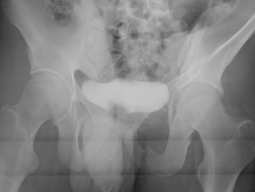
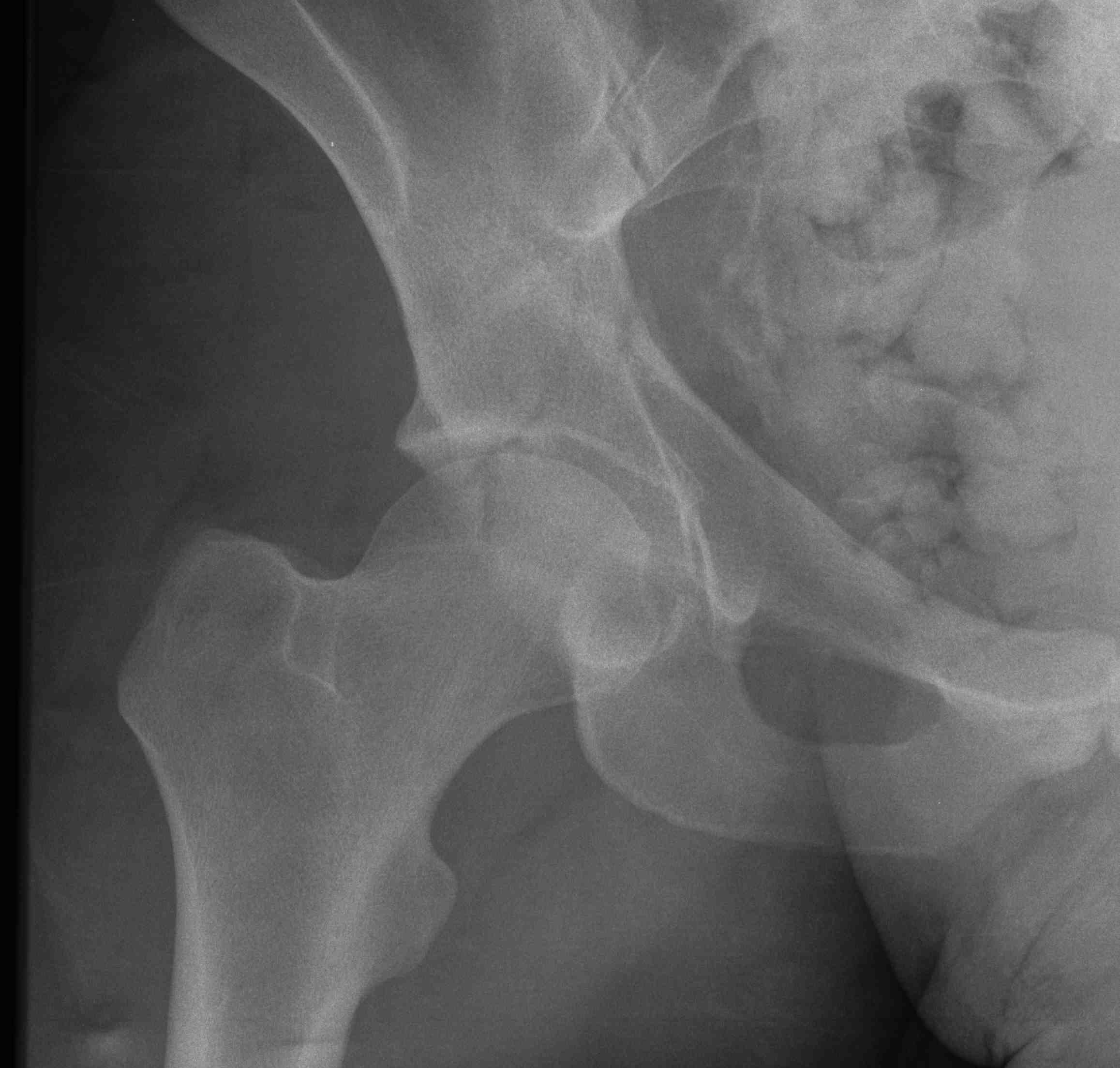
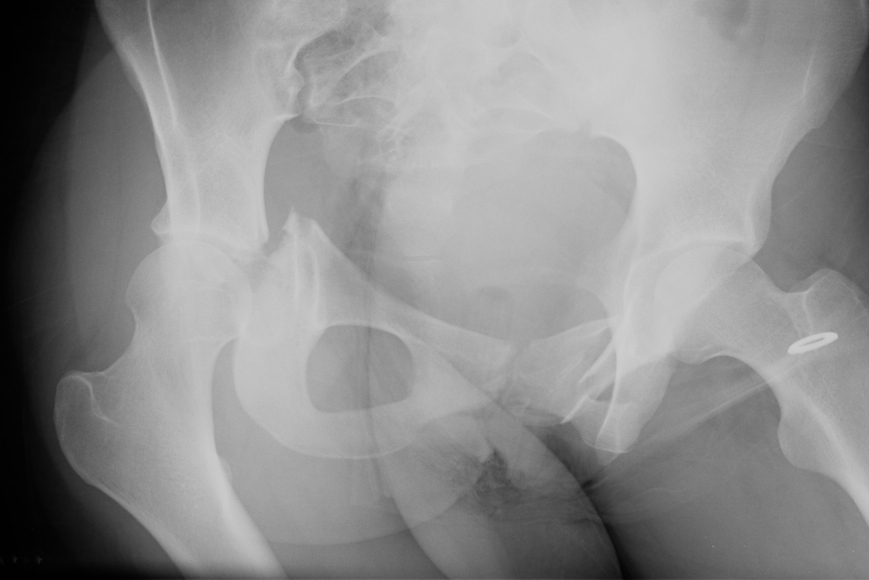
X-ray / 5 standard views
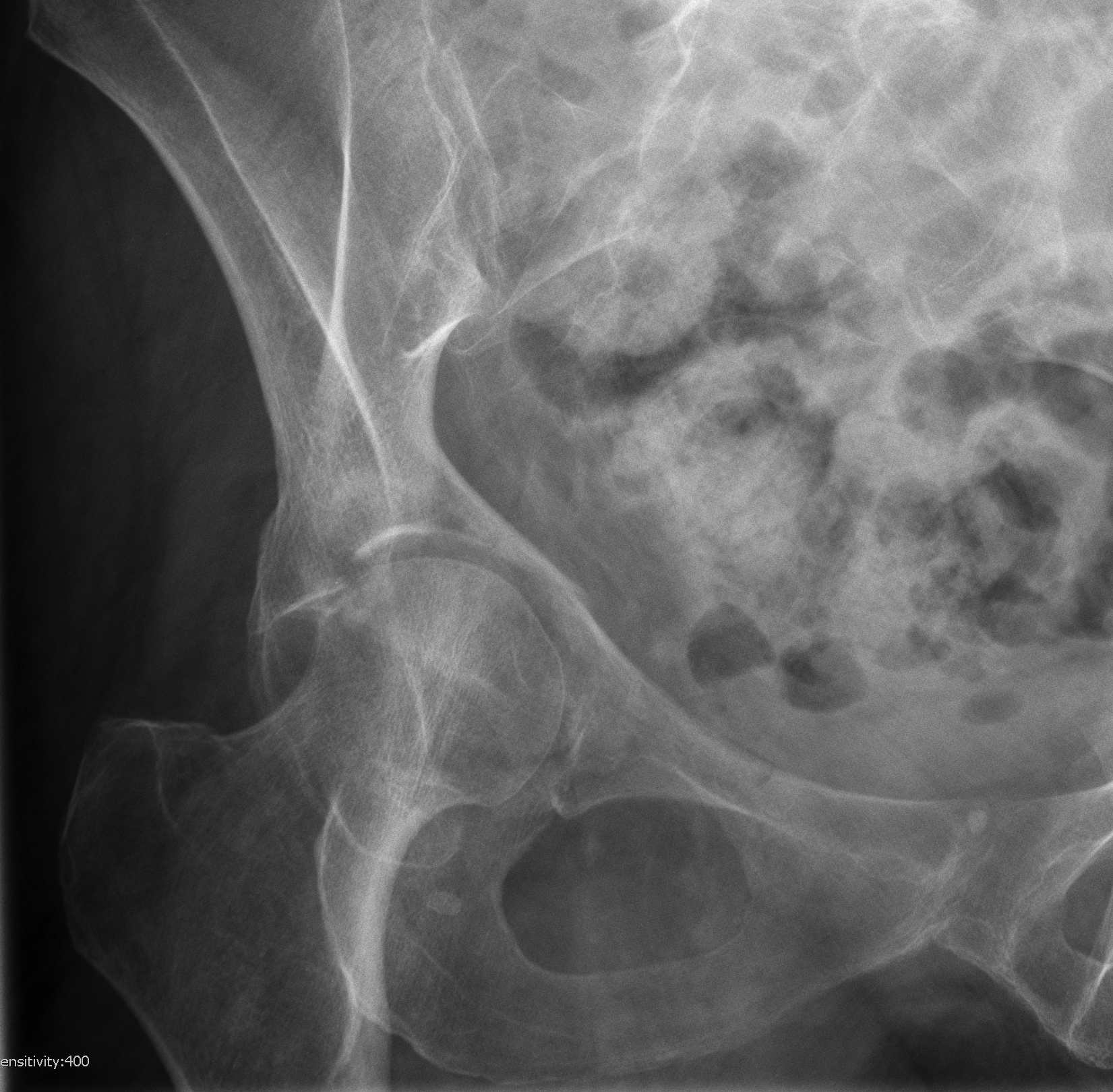
AP / Six X-ray Landmarks
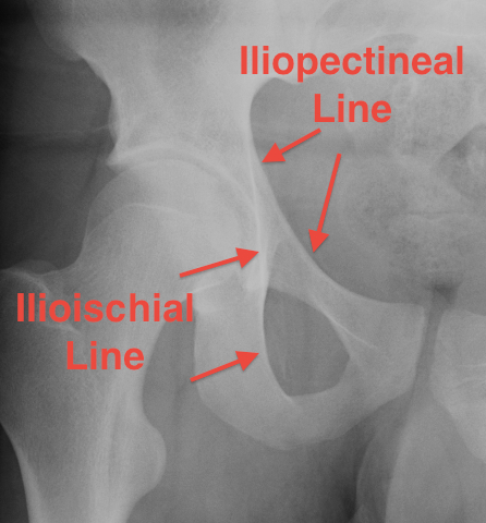
1. Iliopectineal line
- along pelvic brim to pubic symphysis
- anterior column
2. Ilioischial Line
- pelvic brim to ischial tuberosity
- posterior column
- formed by posterior 4/5 of quadrilateral surface ilium
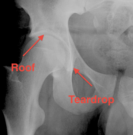
3. The Teardrop
- lateral: subchondral bone condensation at anterior margin of cotyloid fossa
- medial: anterior flat part of quadrilateral surface of iliac bone
4. Roof of acetabulum
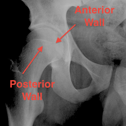
5. Anterior rim of acetabulum
- semilunar
6. Post rim of acetabulum
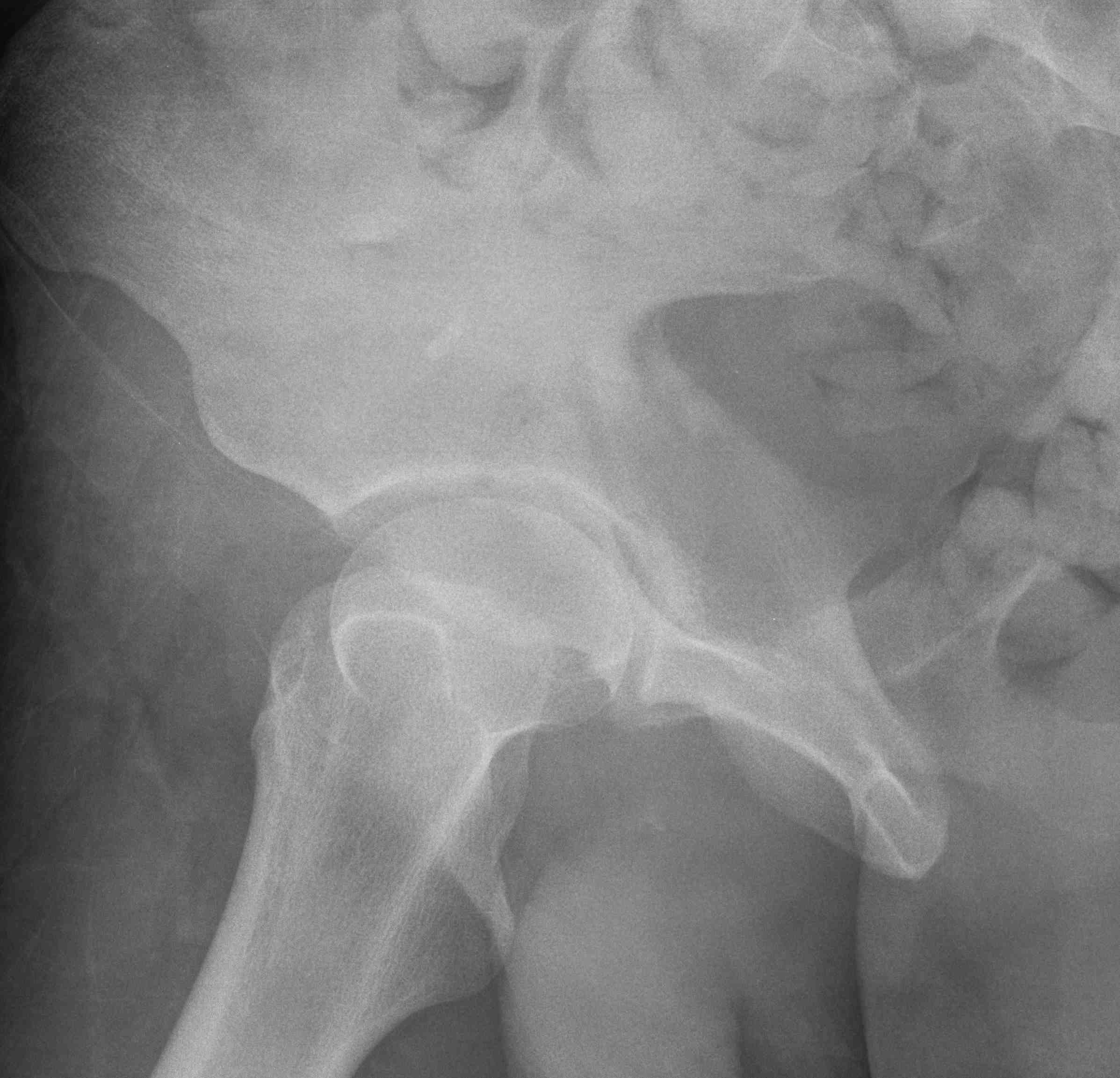
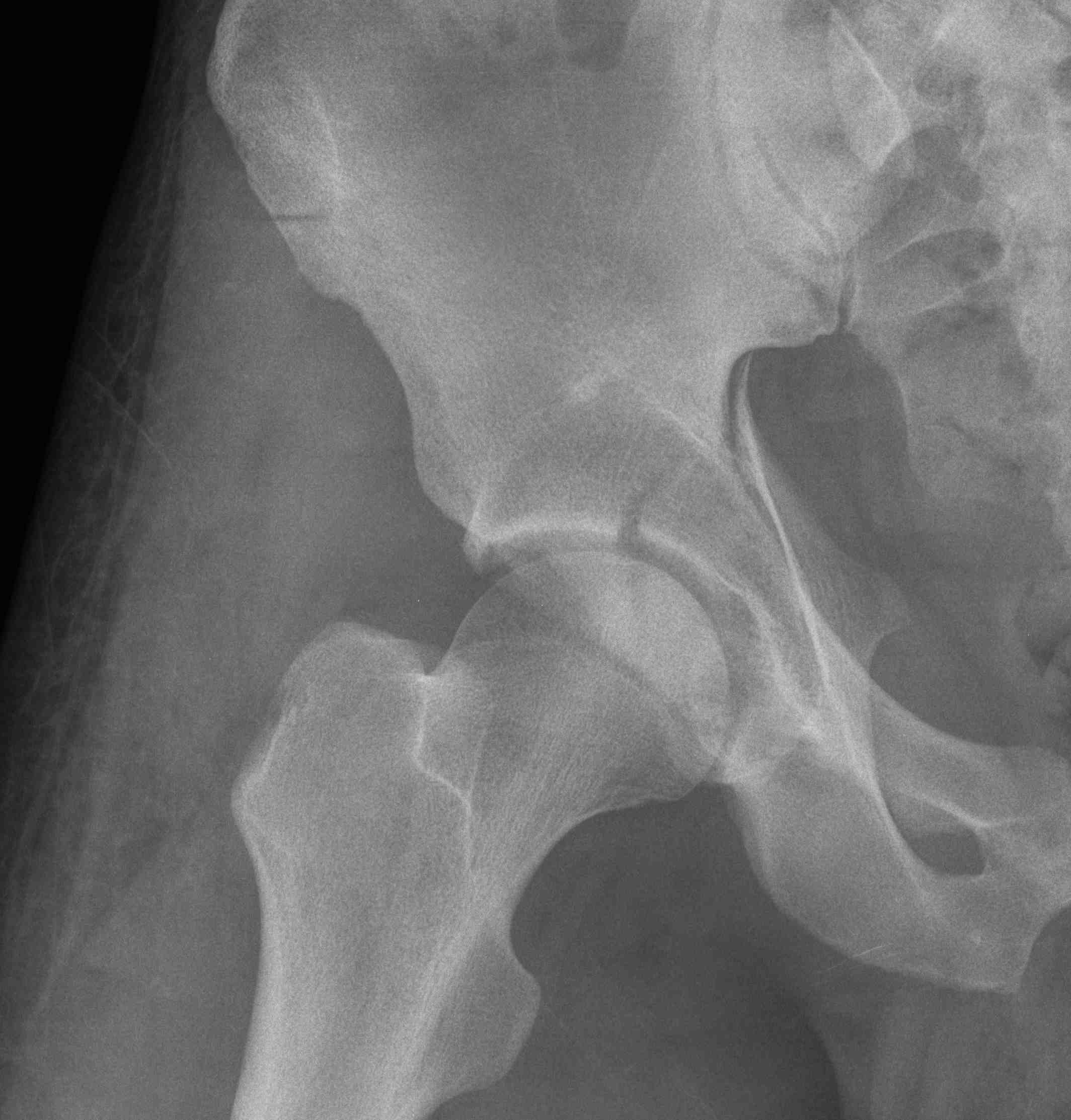
Judet views / 45o obliques
Internal Oblique / Obturator Oblique
- affected side rotated forward
- anterior column + posterior wall
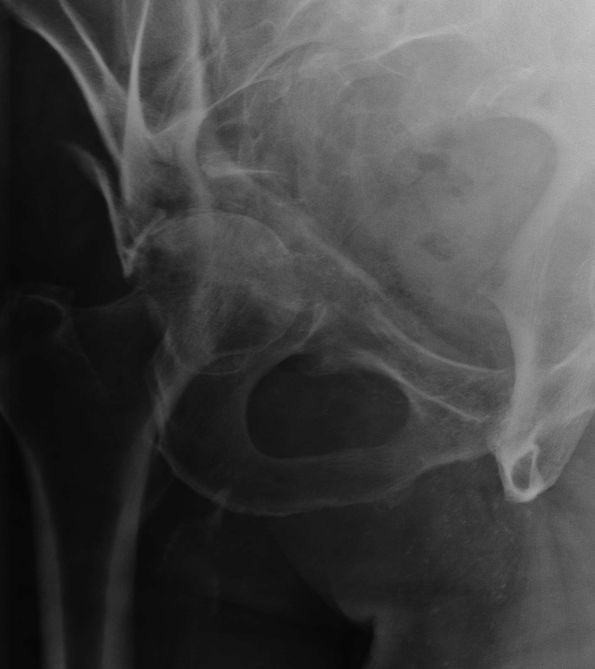
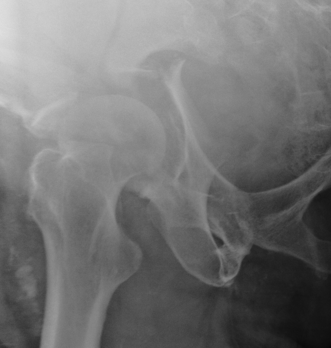
External Oblique / Iliac Oblique
- unaffected side rotated forward
- posterior column + anterior wall
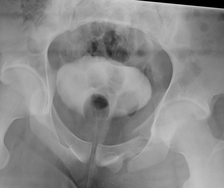
Inlet view / Outlet view
Indicated for pelvic fractures usually
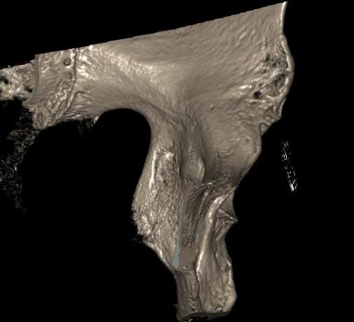
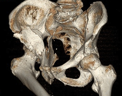
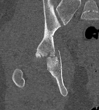
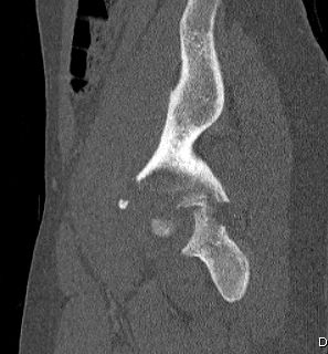
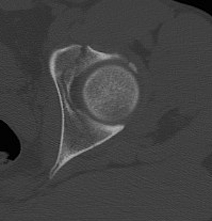
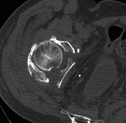
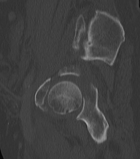
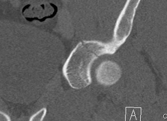
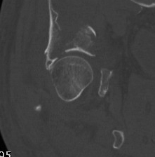
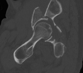
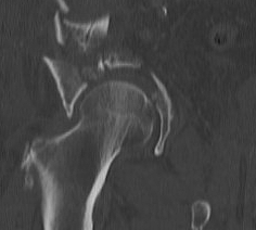
CT
Configuration
1-2 mm sections
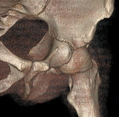
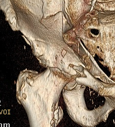
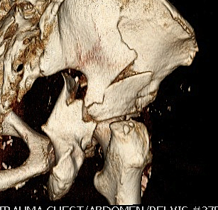
CT reconstruction
- remove head to view acetabulum
- beware volume averaging
- used to guide surgery
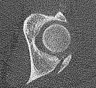
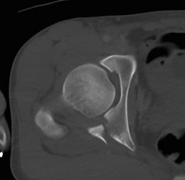
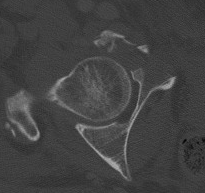
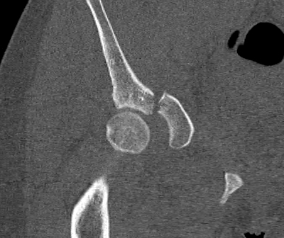
Diagnose
Loose bodies
Femoral head fractures
Subtle subluxation
Articular steps
Roof arc measurement
Letournel Classification
5 Elementary
5 Complex
Elementary / One primary fracture line
1. Posterior Wall
- often associated with posterior dislocation
- may be in one or many pieces
- may have marginal impaction fracture
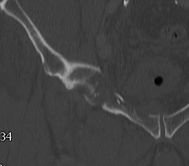
2. Posterior Column
- whole posterior column separated in one piece
- fracture greater sciatic notch
- through inferior acetabulum
- into obturator foramen
- through inferior pubic rami
3. Anterior Wall
4. Anterior Column
- from ilium above ASIS
- through inferior acetabulum
- across obturator foramen
- out through inferior rami
5. Transverse
- from greater sciatic notch to AIIS
- obturator foramen not fractured
High - above acetabulum
Low - through acetabulum
Complex / More than one primary fracture line
1. Posterior column & posterior wall
2. Transverse & posterior wall
3. T-shaped
- transverse through acetabulum
- inferior fracture line to obturator foramen
4. Anterior & posterior hemi-transverse
5. Both column
- Y Shaped transverse above acetabulum
Determinants of outcome
1. Fracture displacement
- < 2mm articular step
2. Fracture location
Early onset of arthritis and poor clinical results correlate with
- displacement present at the time of union within the weight bearing dome
- any roof arc measurement less than 45°
- a broken CT subchondral ring
A. Matta roof arc measurements
Describe location of fracture lines in relation to roof of acetabulum
- integrity of acetabular roof
- must be no hip subluxation
3 roof arc measurements
- AP, 2 Judet's views
- vertical line to centre of head
- line to where fracture enters joint
- the larger the arc, the further the fracture from the roof
- 10o - fracture in roof
- 900 - low fracture
Weight bearing dome is intact if angle > 45o on all 3 views
B. CT subchondral arc
- 10 mm below subchondral bone of roof
- similar to xray roof arc measurements
3. Stability / Concentric reduction
Subluxation
- incongruency between the head and the roof
- poor clinical results are obtained in more than 50% of fractures in which the head is subluxed
- may also have an element of dynamic instability, with certain posterior wall fractures

Any subluxation on CT demonstrates clinical instability
- fractures affecting 40% or more of the posterior wall are usually associated with instability
- fractures less than 40% should be screened for stability under II
4. Other factors
Direct cartilage injury at time of impact
Neurological injury
AVN of head