PIPJ Dislocations
Types
- Dorsal
- Lateral
- Volar
Stabilisers
Proper collateral ligaments
- primary stabilisers
- insert volar third of the base of PP
Accessory collateral ligaments
- inserts on and stabilises lateral margin of volar plate
Volar plate
- thick distally
- thin proximally, allowing collapse during flexion
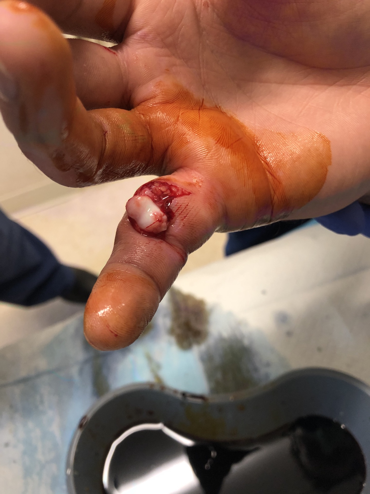
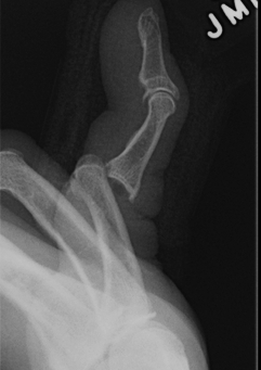
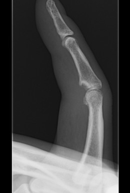
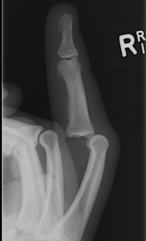
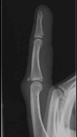
Dorsal dislocations PIPJ
Most common joint injury of the hand
- hinge joint permitting 110o ROM
- volar plate fails distally
- collateral ligaments may be intact
- may be a fracture
Mechanism
- hyperextension
- axial loading of the flexed fingertip
Examination
Stability
- dependant on integrity of the collateral ligaments
- if fragment is > 40 – 50%, the attachment of the true collateral ligament is lost
- unstable
Eaton Classification
I Simple hyperextension
- buddy strap, early ROM
II Dorsal dislocation
- reduced and assess stability
- buddy strap if stable
- extension splint 10o further than instability
- each week extend further by 10o
- early aggressive ROM program
IIIA fracture < 40% volar articular surface
- closed treatment with extension block
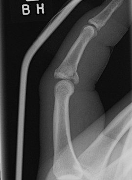
IIIB fracture > 40% + Pilon fractures
- inherently unstable
- extension blocking requires extreme flexion for stability, so risk of flexion contracture is high
- aim for congruent articular surface and early ROM
IIIB Treatment Options
1. Dorsal Blocking K wire
2. Slade Dynamic Distraction External Fixator
3. Compass Hinge
4. Volar Plate Arthroplasty
Dorsal Blocking K wire
Technique
- flexion P2
- dorsal entry into P1
- 40o flexion
- early removal at 3/52
- Improvement compared to extension blocking
Suzuki / Slade Dynamic Distraction external fixator
Concept
- closed reduction through ligamentotaxis
- early motion of PIPJ
Technique
- transverse K wire in rotational centre / head P1
- transverse K wire distal P2
- attached by rubber bands
- third K wire mid-diaphysis P2, prevents dorsal translation of MP
Deshmuhk S etal JBJS Br July 2004
- 12 patients complex fracture dislocations PIPJ
- treated with modified pin / rubber band system
- average 84o ROM
- nil radiological osteolysis or clinical osteomyelitis
- all returned to occupation
Hotchkiss designed PIP compass hinge
Technique
- K wire to centre head of P1 to set centre rotation
- 2 x K wires each in P1 / P2
- barrel over centre of rotation
- options of active motion, passive ROM, locked
Bain I JBJS Br 1998
- 12 patients
- mean range of motion 12 – 86o
- only half presented within 2 week of injury
- combined operation with ORIF and volar plate arthroplasty
- nil osteomyelitis
- hinge on for 6 weeks
Volar plate arthroplasty / Volar plate advancement
Technique
- incise accessory collaterals to release volar plate
- excise bony fragment
- suture proximal volar plate into defect
- pass sutures through drill holes in base P2
- tie over button dorsally
- dorsal blocking splint 4 - 6 / 52
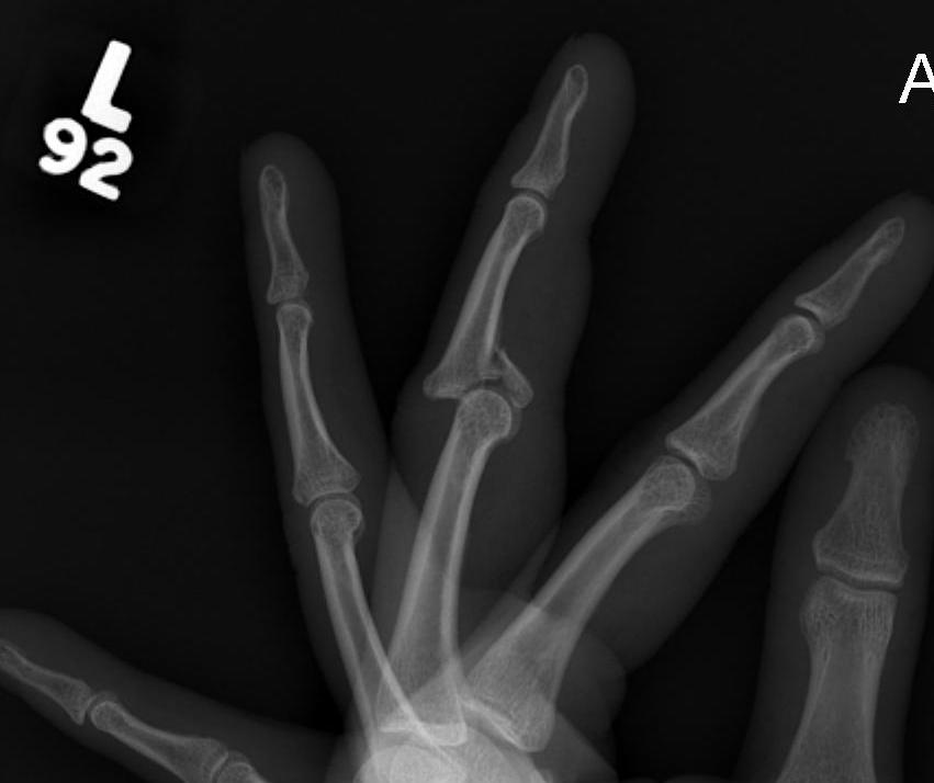
Volar PIPJ dislocations
A. Straight volar dislocation
Assessment of central slip post reduction critical
- if can active extend to within 30o, splint extended
- if nil active, surgical repair to prevent boutonniere
B. Volar rotary subluxation
- condyle button holes between central slip and lateral band
- irreducible dislocation
Lateral PIPJ dislocations
Rupture of one collateral ligament and volar plate
- may be bony avulsion
Management
- reduce and hold in extension 2/52, then protected ROM
- can perform primary repair or reconstruct