Types
Anterior ankle arthroscopy
Posterior ankle and hindfoot arthroscopy
Anterior ankle arthroscopy
Indication
Osteochondral lesions
Loose bodies
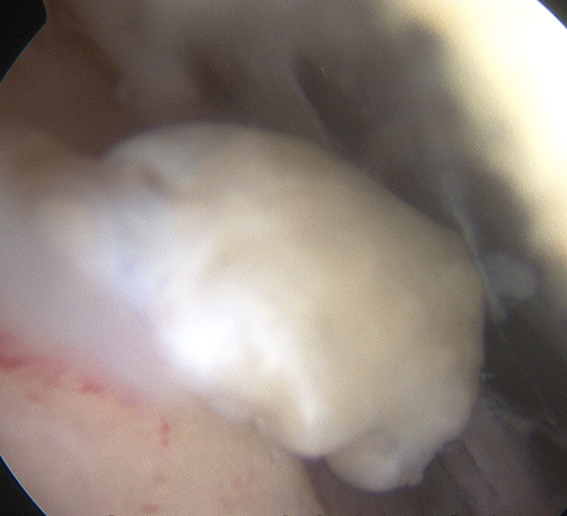
Tibial talar anterior impingement
Syndesmotic injuries
Anterolateral impingement
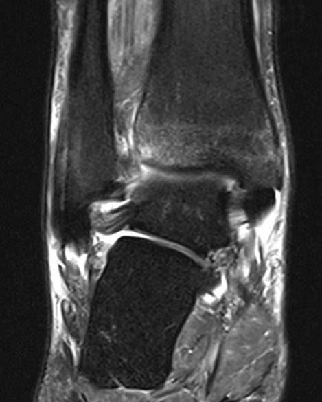
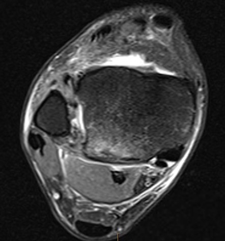
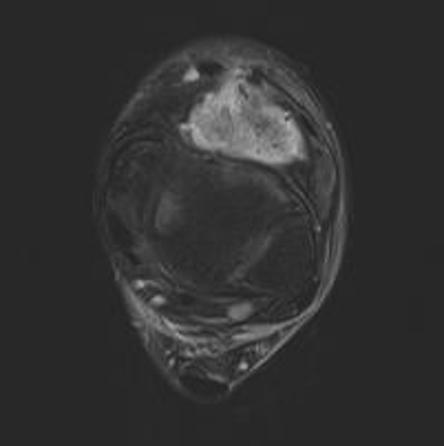
PVNS
Ankle arthrodesis
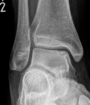
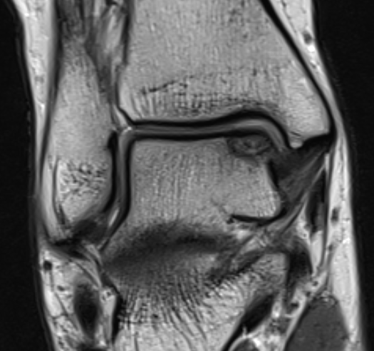
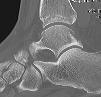
Talus ostechondral lesions
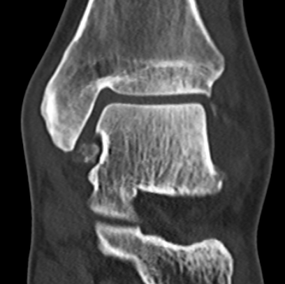
Ankle loose bodies
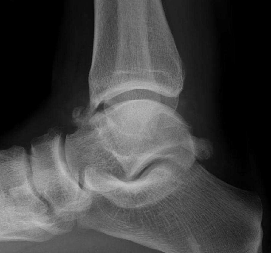
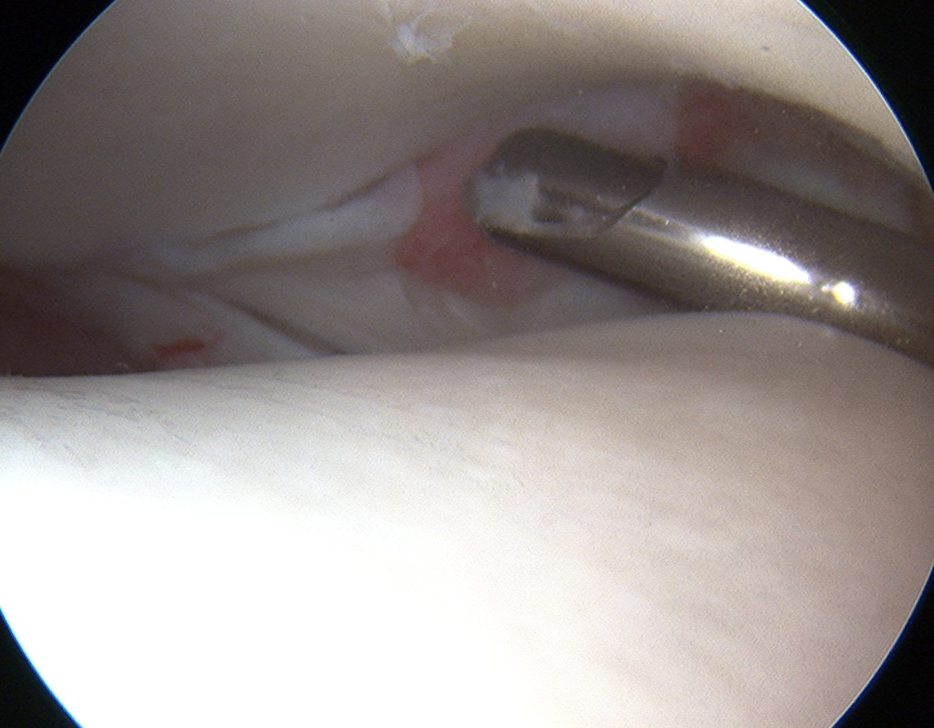
Anterior tibial spur and tibio-talar impingment
Position
Supine on table with tourniquet +/- traction
- standard knee arthroscope which allows outflow
- 2.7 mm 30° scope - smaller but no outflow
- often insufflate ankle joint with fluid prior to portals
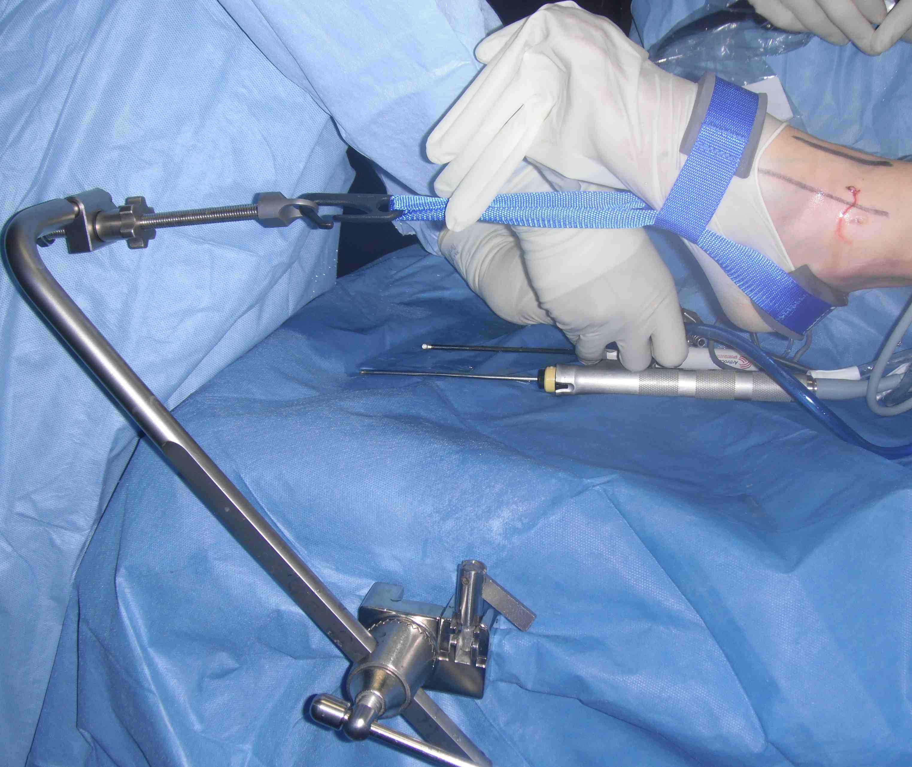
Traction
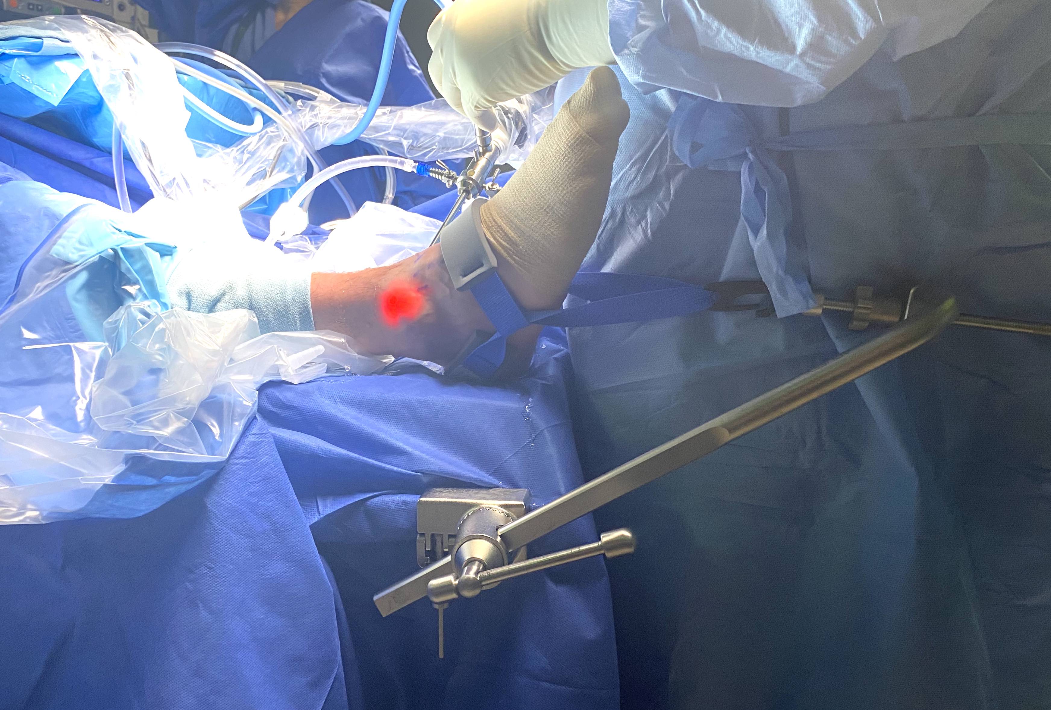
Foot halter traction attached to bed
Options
Halter traction - to bed / to surgeon
Calcaneal pins
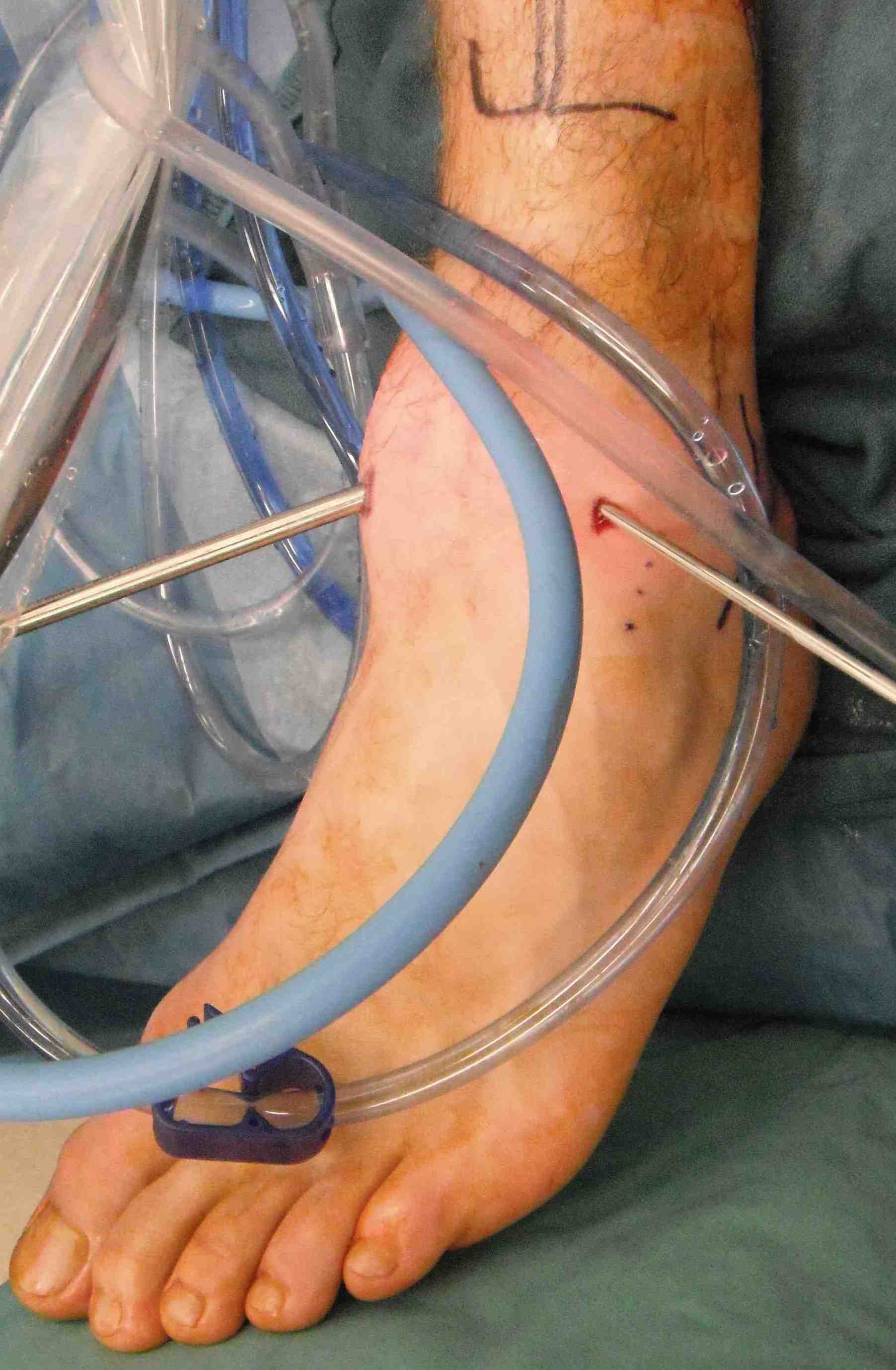
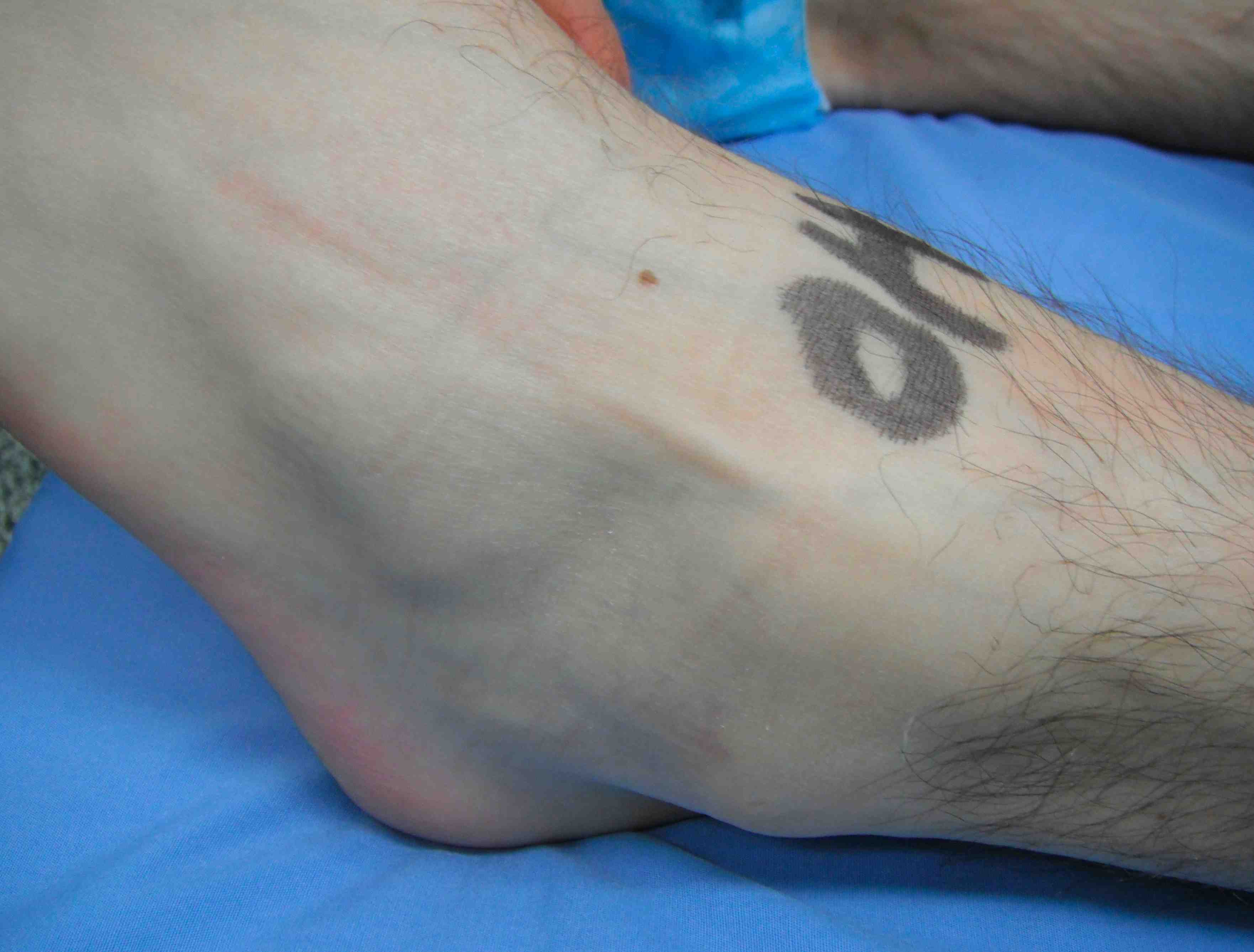
Portals
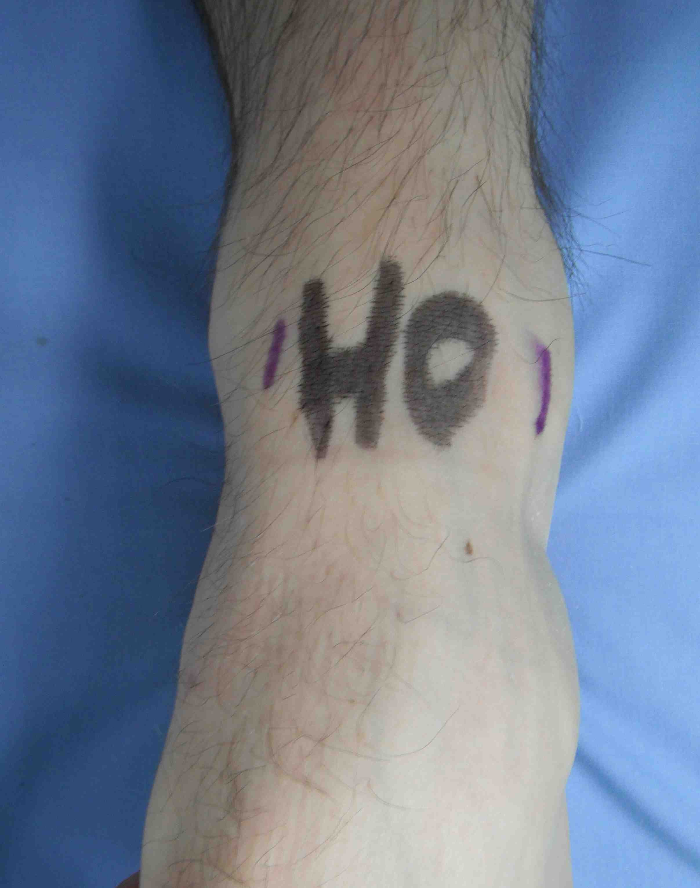
Anteromedial and anterolateral portals
Anterolateral (AL) portals
- lateral to peroneus tertius
- often inserted first so can transilluminate and avoid saphenous nerve on AM portal
- structure at risk is branches superficial peroneal nerve
- incision in skin only and blunt dissect down to capsule
- insert blunt trochar behind anterior extensor tendons aiming medially
Anteromedial (AM) portal
- medial to tibialis anterior
- structure at risk is great saphenous vein and saphenous nerve
- use transillumination to avoid
- insert and visualise needle
- skin incision only and blunt dissection to capsule
Superficial peroneal nerve
Technique
Vumedi 21 point ankle arthroscopy video
Arthroscopy techniques ankle arthroscopy
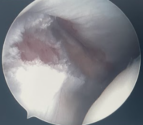
Inspect
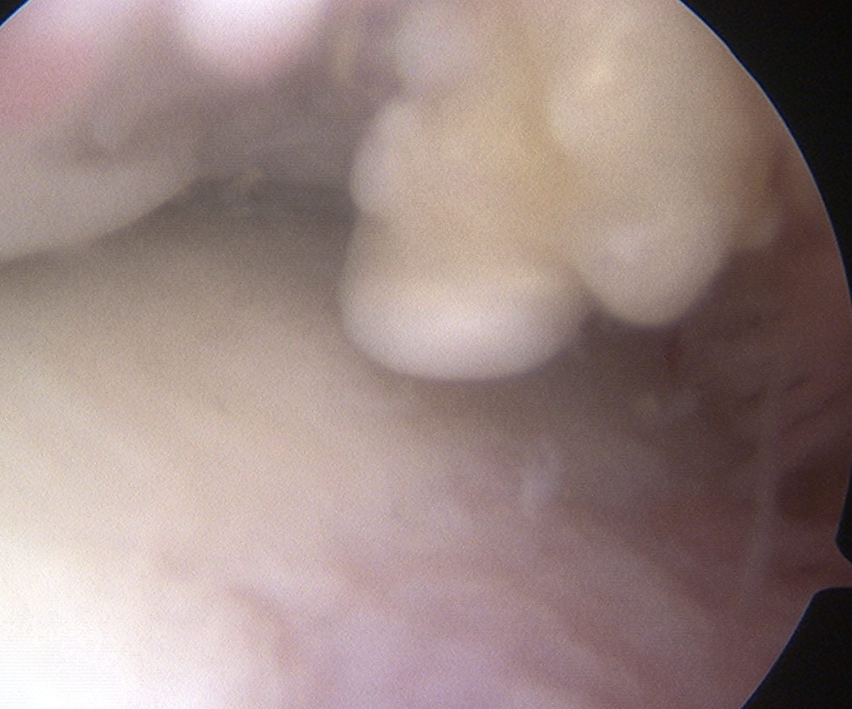
- talar dome and tibial plafond for chondral lesions
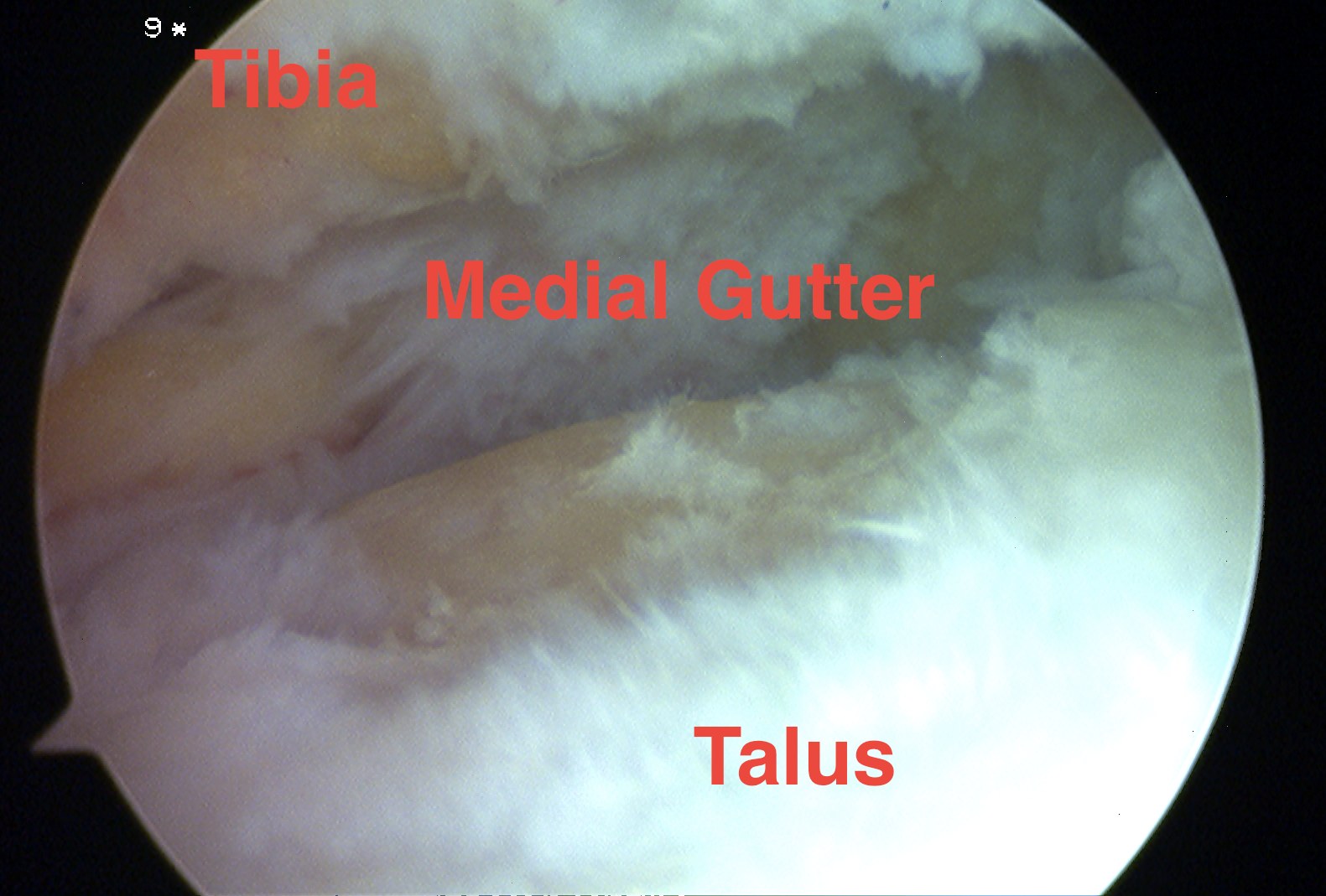
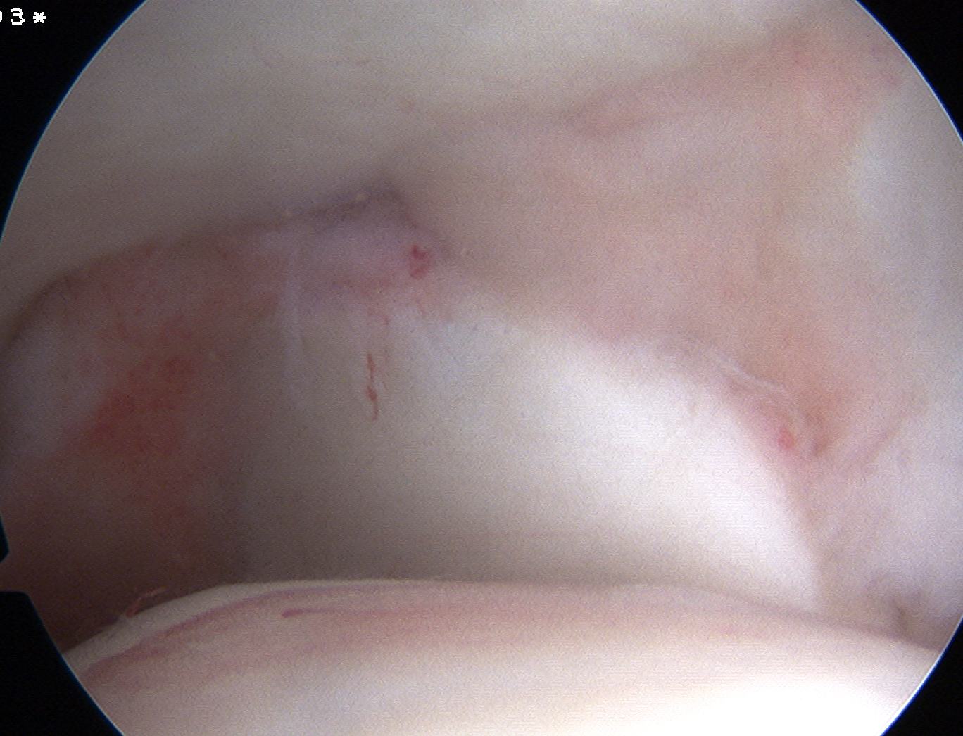
- medial gutter
- lateral gutter
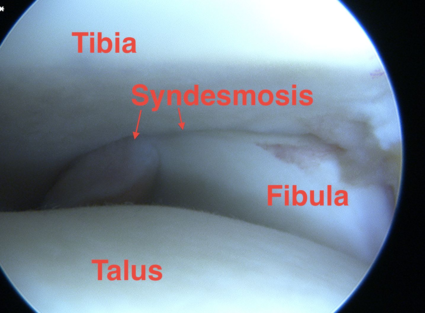
- syndesmosis
- posterior capsule and FHL
Talar dome
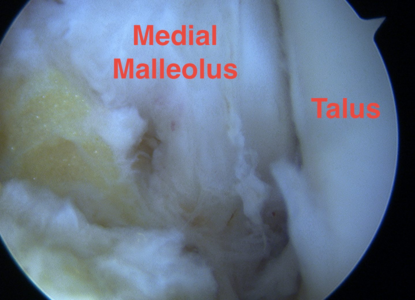
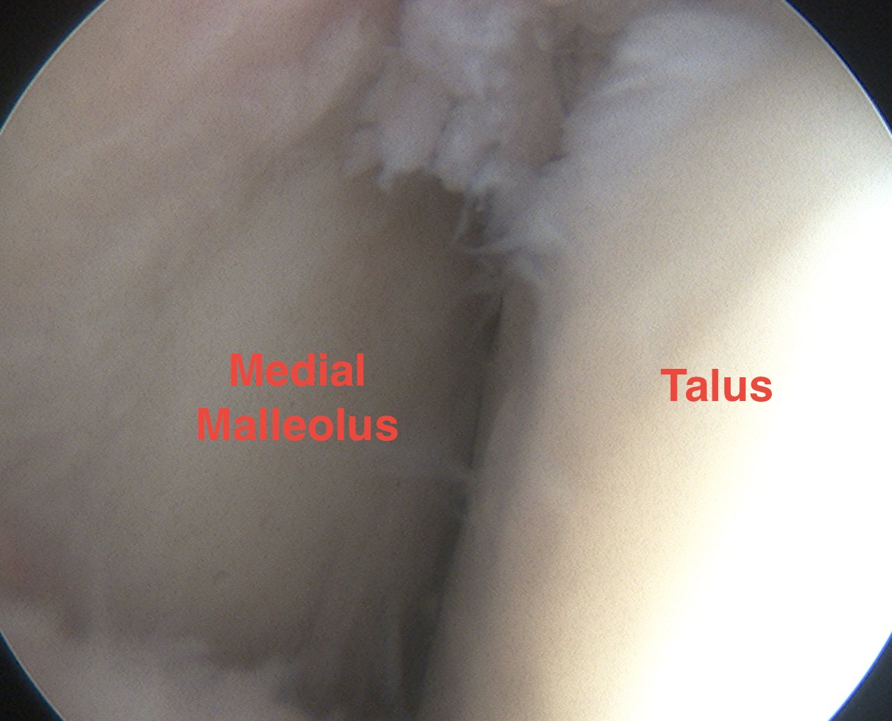
Medial gutter
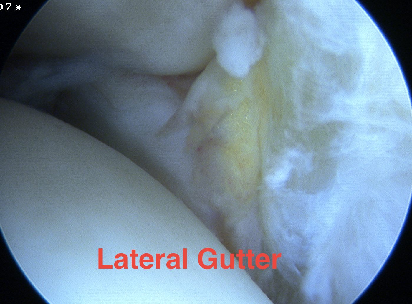
Lateral gutter
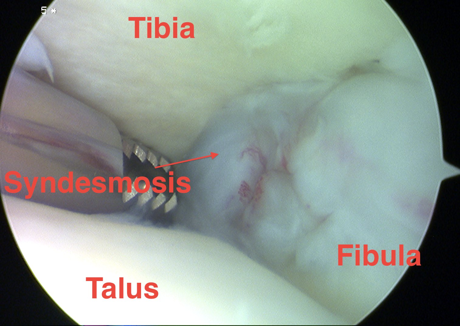
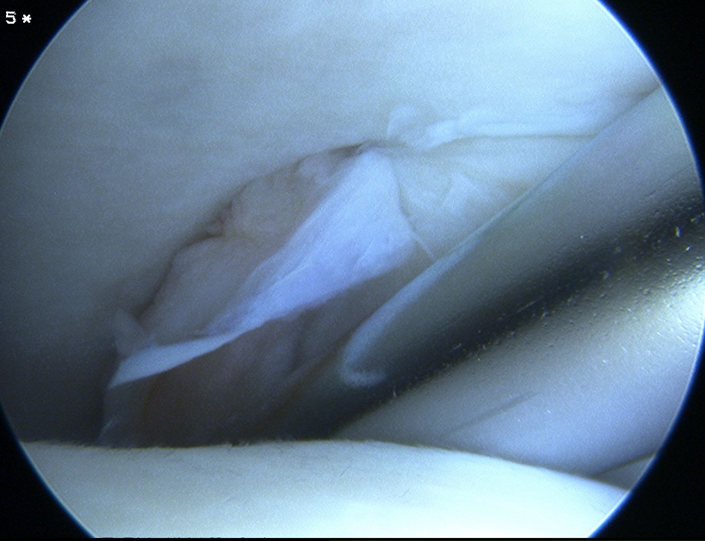
Syndesmosis
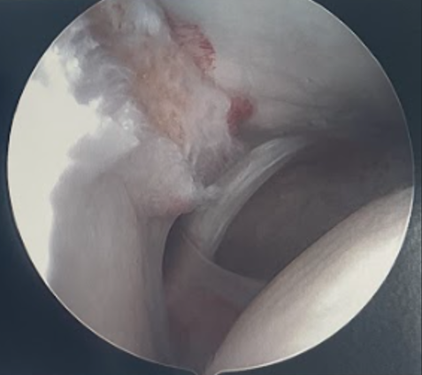
Disrupted and unstable syndesmosis
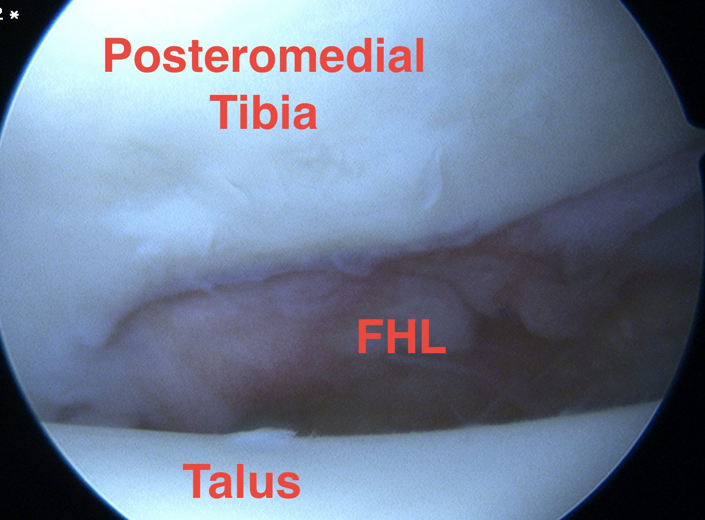
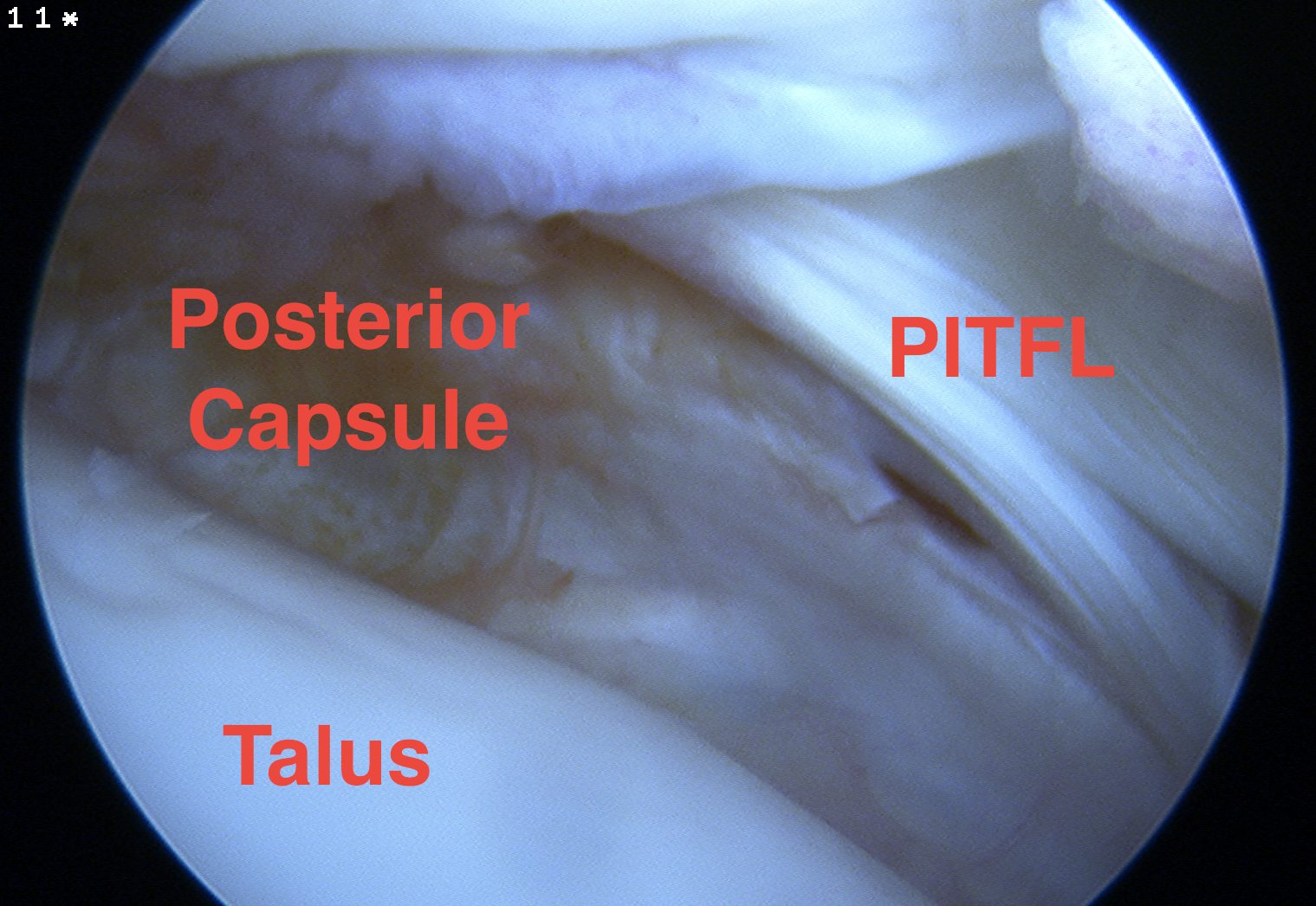
Posterior capsule and FHL
Complications
- systematic review of complications following anterior ankle arthroscopy
- overall complication rate 4%
- neurological injury 2%
- superficial peroneal nerve most commonly injured
- some deep peroneal / saphenous
Rare
- compartment syndrome - extravasation of fluid into calf
- infection
- DVT
- pseudoaneurysm

Pseudoaneurysm following anterior ankle arthroscopy
Posterior ankle and hindfoot arthroscopy
Indications
Insertional achilles tendinosis / Haglund's deformity / retrocalcaneal bursitis
Posterior ankle impingement - os trigonum / FHL tendinopathy
Posterior talus osteochondral lesions
Subtalar arthritis / arthrodesis
PVNS
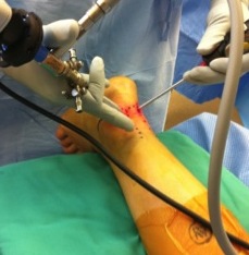
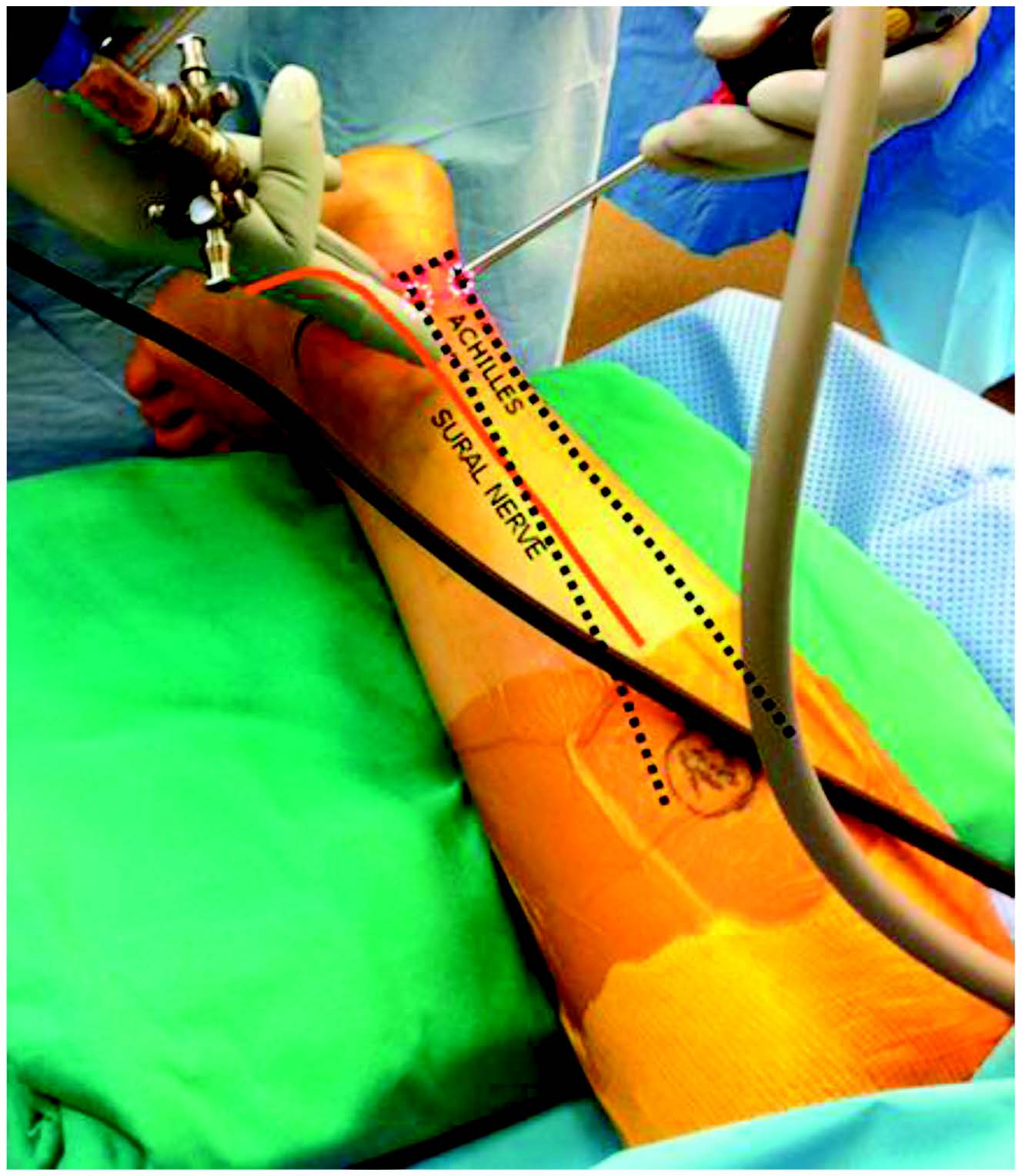
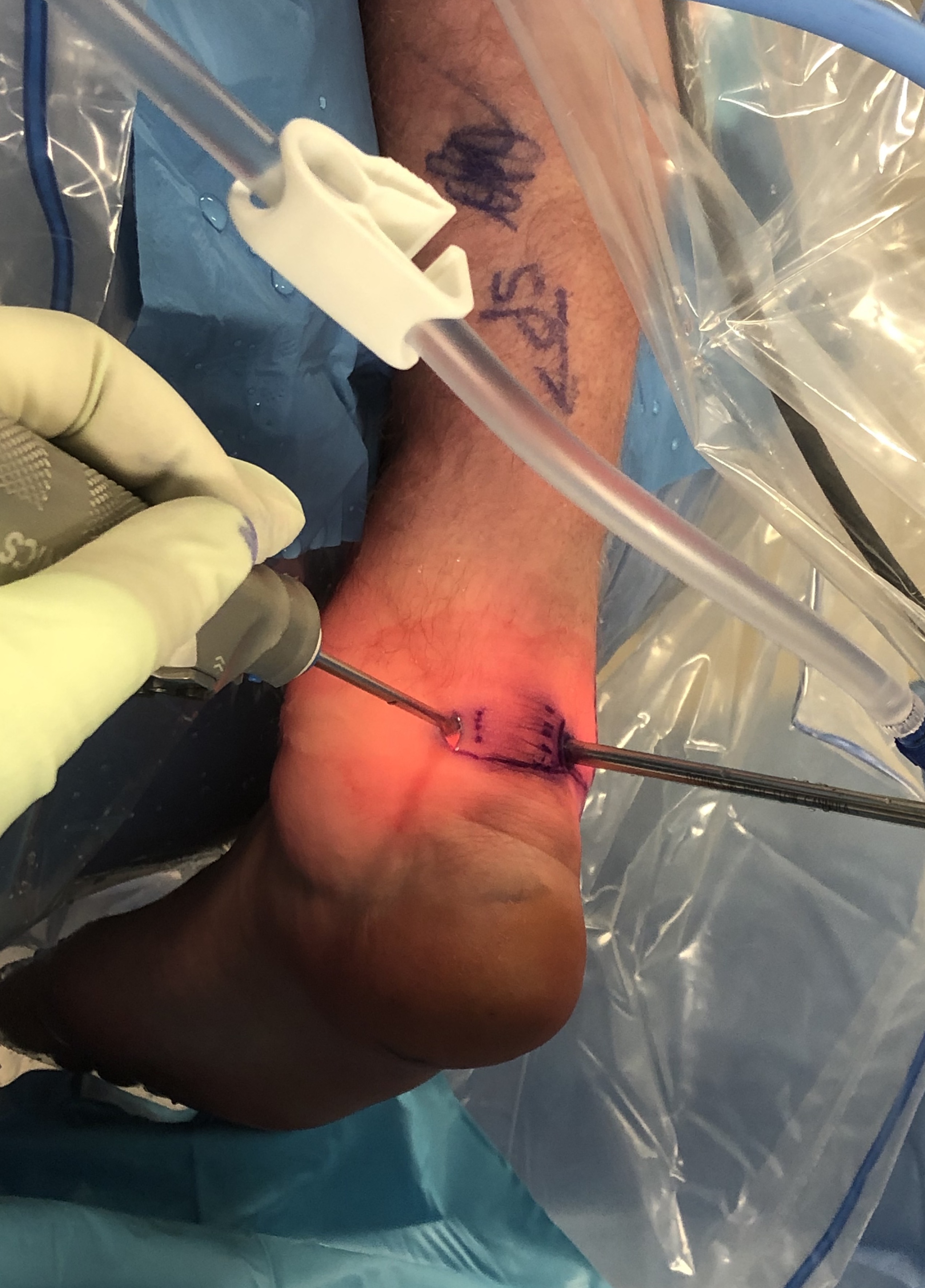
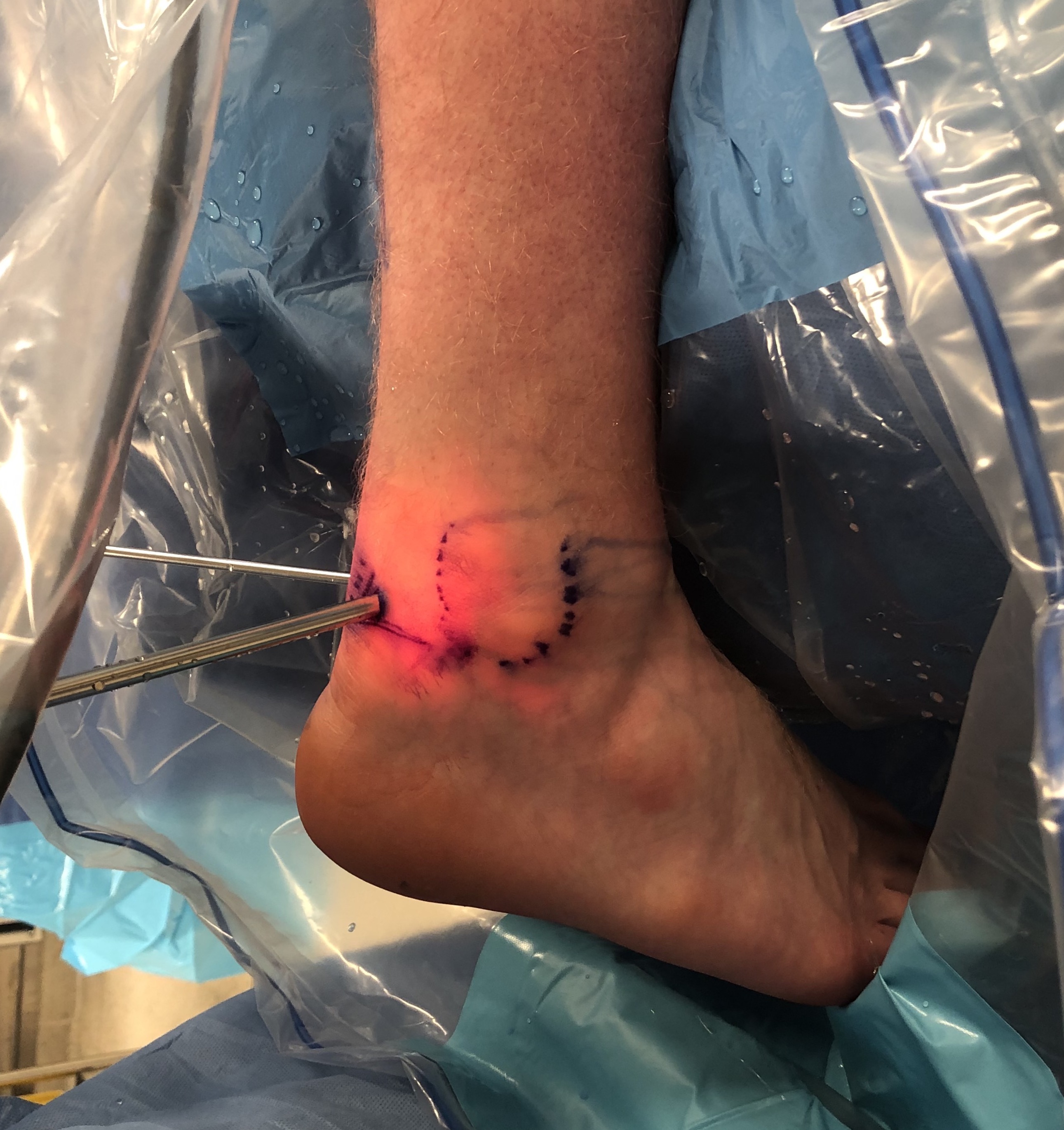
Portals
Posterolateral
- just lateral to tendoachilles tendon
- at the level of or just above the tip of fibula
- aim for 1st webspace
- stay medial to sural nerve
Posteromedial
- just medial to tendoachilles tendon
- at the level or just above the tip of the medial malleolus
- aim laterally to avoid posteromedial neurvascular structures
- aim for webspace between 2nd and 3rd toe
Technique
Vumedi posterior ankle arthroscopy portals
Vumedi posterior ankle arthroscopy technique
JBJS Essential Surgical Techniques PDF
Complications
- systematic review of complications following posterior ankle arthroscopy
- overall complication rate 7%
- neurological injury 4%
- 189 posterior ankle and hindfoot arthroscopies
- 2% sural nerve injury, 2% plantar numbness
- 1% infection