MRI

Achilles Tendonopathy

Definition


Inflammation of achilles tendon; insertional or noninsertional


Spectrum


Tendonitis / Tendonosis / Rupture
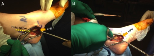
 

Anatomy


Triceps surae

- medial and lateral gastrocnemius

- soleus

- surrounded by paratenon which allows gliding and supplies nutrition


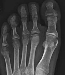
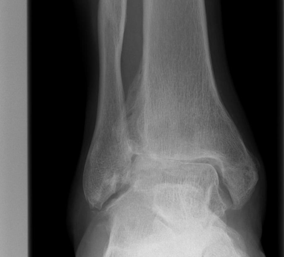
Inserts middle 1/3 calcaneal tuberosity

- 2 x 2 cm area

- 90o rotation distally


Retrocalcaneal bursa (x2)

Plantar Fasciitis

Definition


Pain at attachment of thickened central part of plantar aponeurosis to Medial Calcaneal Tuberosity


Anatomy Plantar Fascia


Origin 

- medial calcaneal tuberosity


Inserts 

- 5 bands superfical & deep layers


Superficial

- insert transverse MT ligament & skin


Deep 

- flexor sheath, volar plate & periosteum of P1


Tendonitis / Tendonopathy

Anatomy


Lateral compartment of leg

- run through retromalleolar groove

- pass superior and inferior to peroneal tubercle

- covered by inferior peroneal retinaculum


Peroneus longus

- origin lateral condyle of tibia and head fibula

- tendon PL superficial and inferior to brevis in retromalleolar groove

- runs in cuboid groove 

- insert plantar surface base of 1st MT and lateral aspect medial cuneiform

Freiberg's

Definition


Crushing osteochondritis of metatarsal head


Frieberg's


Epidemiology


Usually 2nd metatarsal (80%)

- occasionally third

- can occur in any


Age 10-15 years

- peak 15 year old girls

- F:M = 3:1

- occurs during the growth spurt at puberty


Bilateral in 6%


Aetiology


Ankle Osteoarthritis

AetiologyAnkle OA


Trauma


A. Ankle Fracture
 

Types

- Weber A 4%
- Weber C 33%
- Displaced large posterior malleolar


Any OA develops in first 2 years


Causes

- articular damage at time of injury
- non anatomical reconstruction
- complications i.e. infection


Thoraco Lumbar Fracture

Xray Assessment


A:  Alignment

B:  Bony

C:  Canal

D:  Disc

S:  Soft tissues


Goals of surgery


1.  Correct deformity

2.  Restore stability

3.  Decompress neural elements if required


MRI


Advantage

- defines level of conus

- may need anterior rather than posterior surgery if lesion above conus

Lumbar Stenosis

DefinitionLumbar Stenosis


Reduction of space available for neural elements in spinal canal or intervertebral foramina

- due to degenerative changes, congenital abnormalities or both

- involves compression of the thecal sac or nerve roots


Epidemiology


Onset 50 - 60's

- M = F

- associated with onset OA spine

Thoracic Disc Disease

Epidemiology


0.05% incidence

- rare due to stabilising effect of rib cage

- even more rare to have symptoms


Reasoning

1.  Discs are narrower

2.  Foramina larger

3.  Thoracic spine

- facet joints orientated for rotation

- lumbar spine for flexion extension

- flexion is typically the motion which ruptures annulus


Anatomy