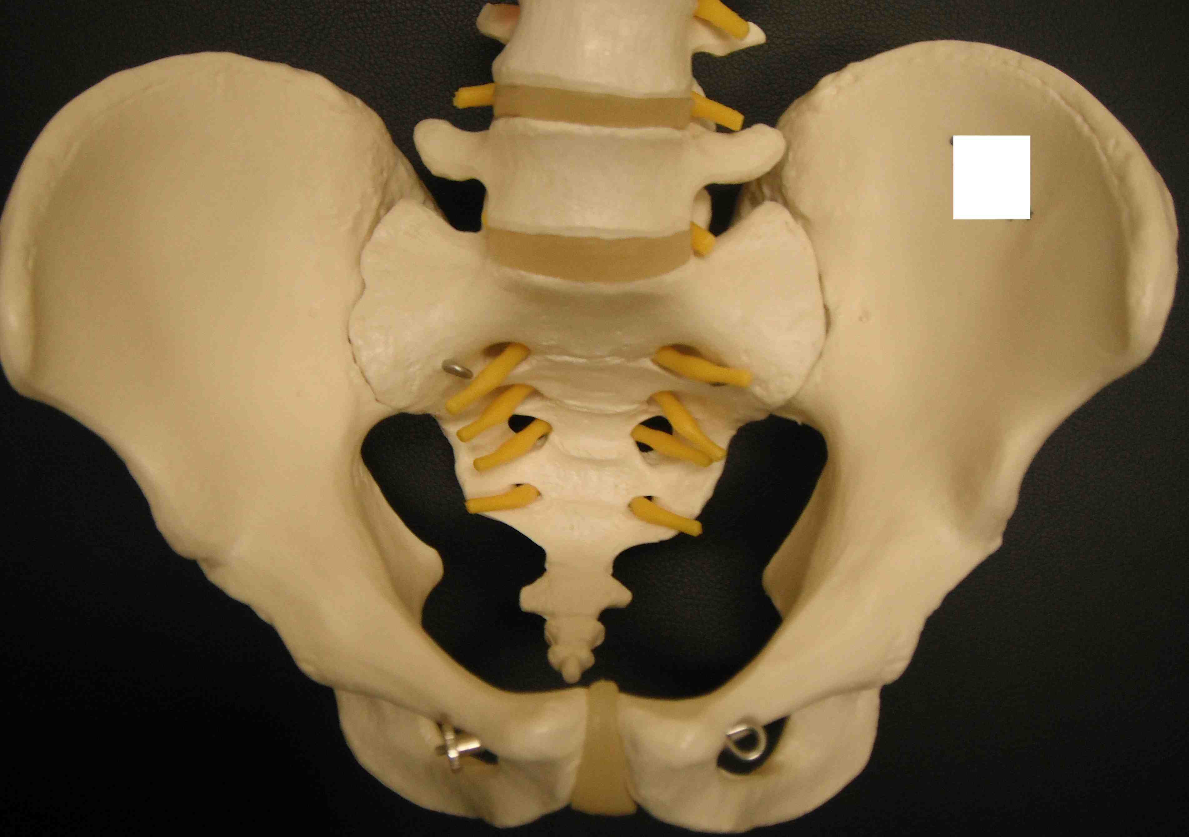
Bony anatomy
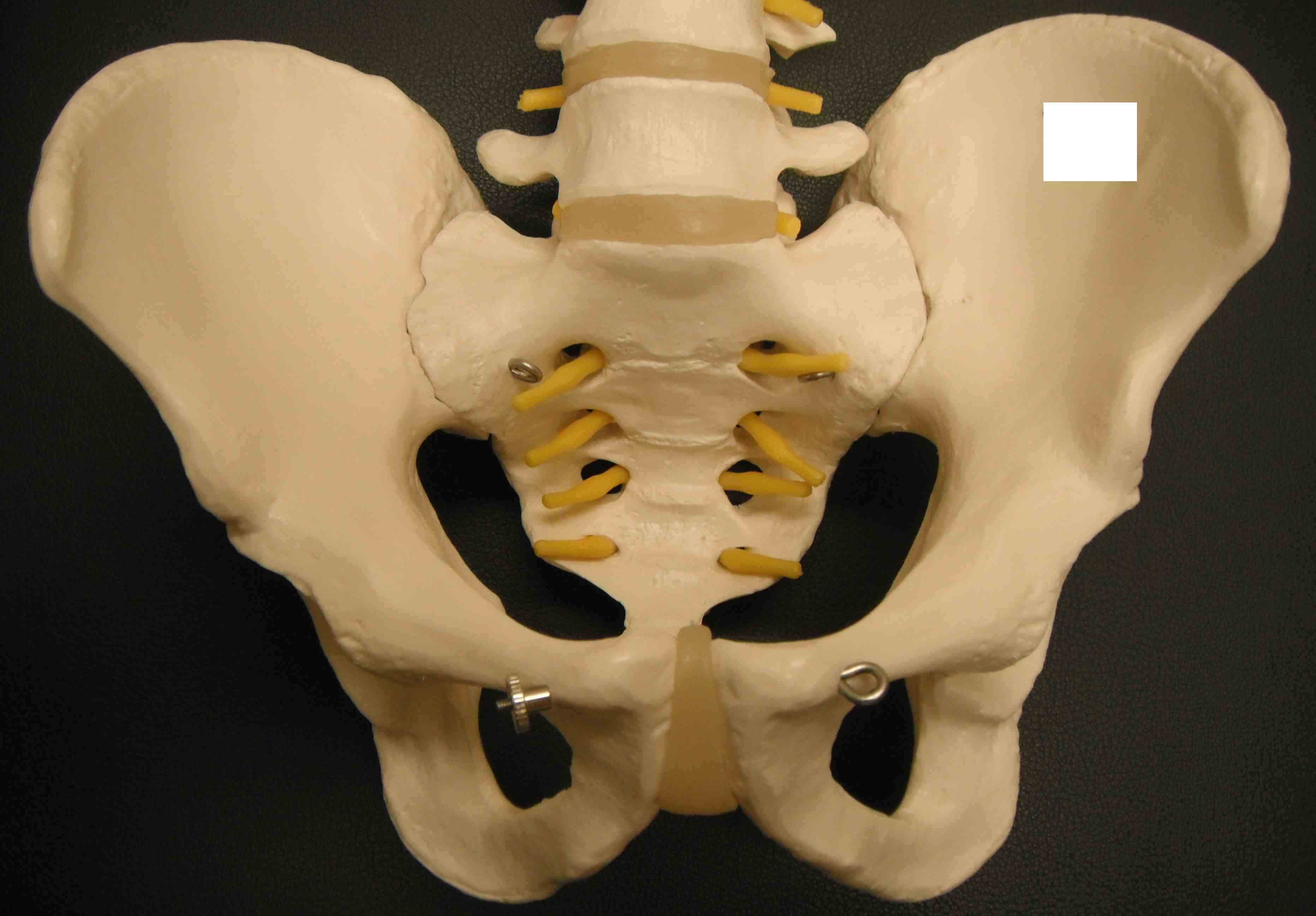
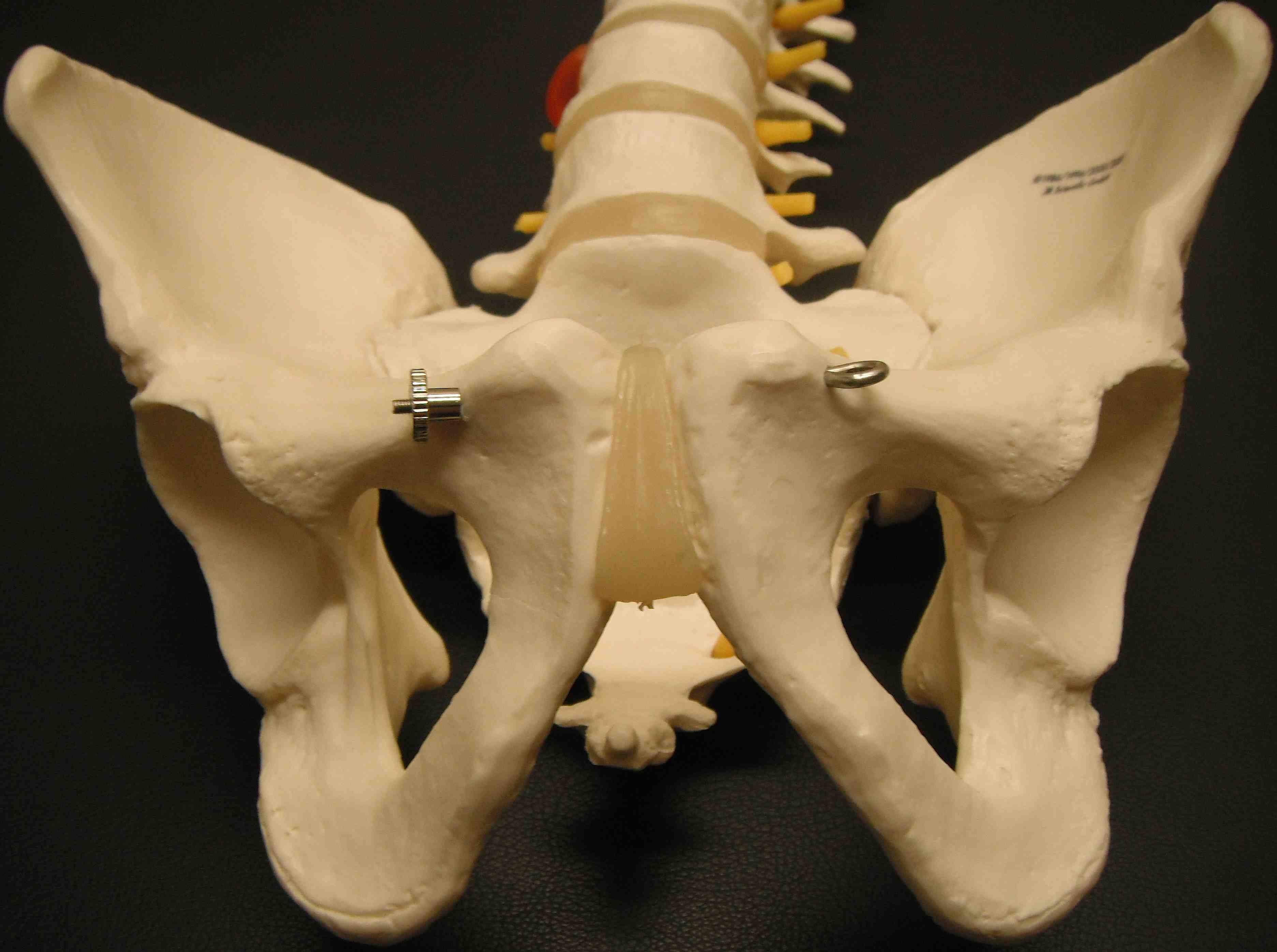
Soft tissue anatomy
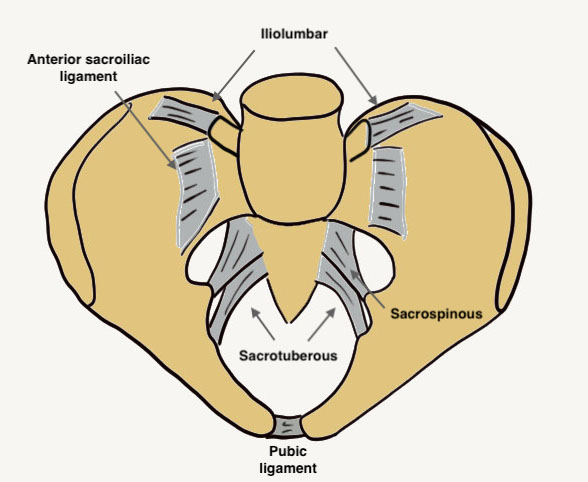
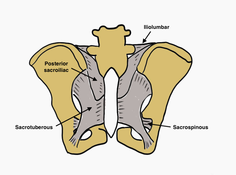
| Anterior sacroiliac ligaments | Resist external rotation |
| Posterior sacroiliac ligaments | Strongest in the body |
| Sacrospinous ligaments |
Lateral sacrum to ischial spine Resist external rotation |
| Sacrotuberous ligaments |
Posterior sacrum to ischial tuberosity Resist vertical shear |
| Iliolumbar ligaments | Iliac crest to transverse process of L5 |
Young and Burgess Classification
|
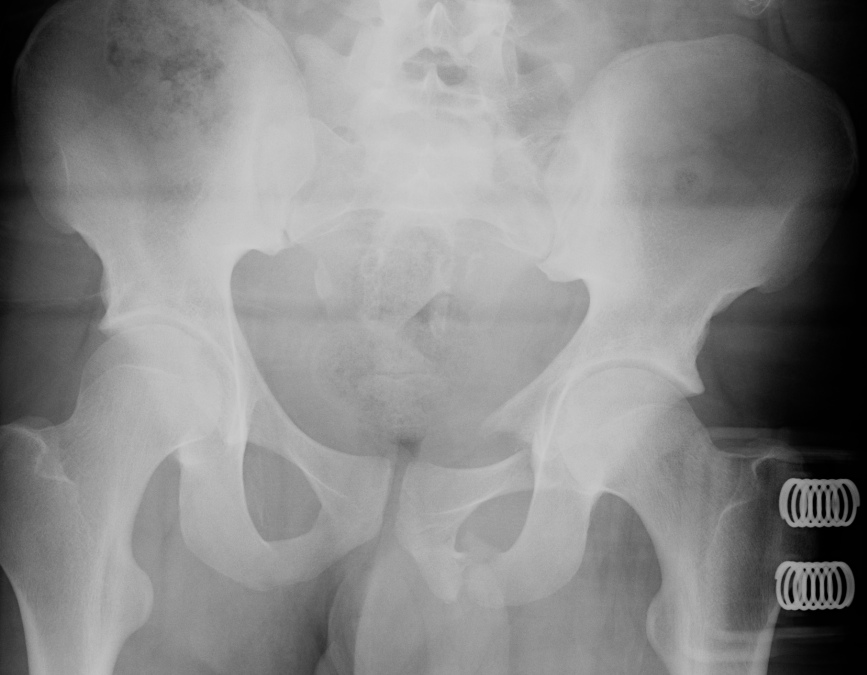
AP COMPRESSION Diastasis of the pubic symphysis without anterior fracture |
APC 1 | Pubic diastasis < 2.5 cm | Stable |
| APC 2 |
Pubic diastasis > 2.5 cm Anterior SI joint widening Posterior SI ligaments intact |
Rotationally unstable Vertically stable |
|
| APC 3 |
Pubic diastasis > 5 cm Anterior and posterior SI joint widening |
Globally unstable
|
|
|
LATERAL COMPRESSION Transverse overlapping obturator ring fractures |
LC1 | Sacral impaction fracture | Stable |
| LC2 | Iliac wing fracture |
Rotationally unstable Vertically stable |
|
| LC3 |
Lateral compression fracture one side AP compression fracture other side |
Globally unstable | |
|
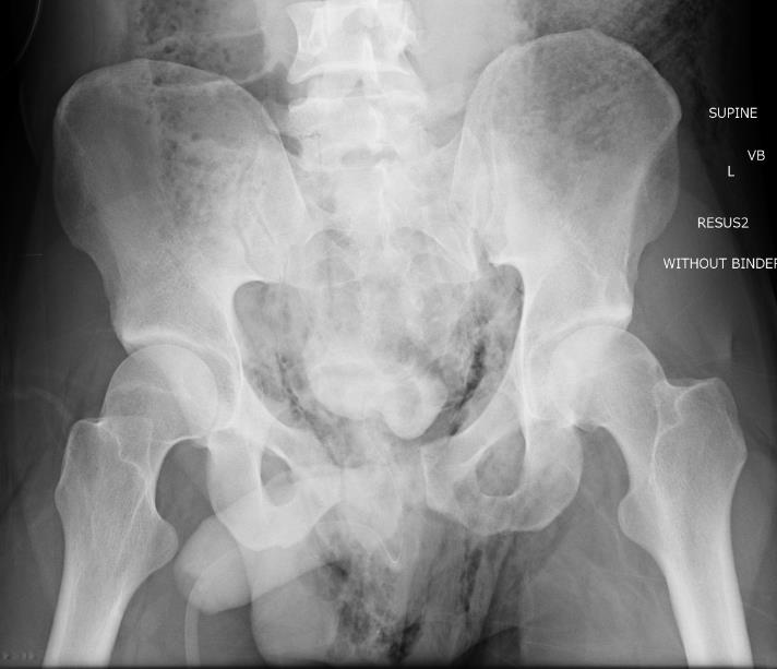
VERTICAL SHEAR FRACTURE Fractures of the pubis and SI joint with vertical displacement |
Vertical displacement of hemipelvis Fractures of pubis and SI joint |
Unstable | |
|
COMBINED |
Complex fractures with combined elements of ACP / LC / Vertical shear |
|
APC / Anterior Posterior Compression
APC-1
< 2.5 cm diastasis with no anterior SI joint widening
APC-2
Pubic diastasis with anterior SI joint widening on the right
APC-3
Pubic diastasis > 5 cm with complete SI joint disruption
LC / Lateral Compression
Mechanism
Compressive force to lateral aspect of the pelvis
Results in internal rotation and medialisation of the hemipelvis
LC-1
Pubic rami + sacral compression left side
LC-2
Pubic rami + iliac wing fracture
LC-3
Wind swept pelvis
Lateral compression + contralateral open book
Vertical Shear
APC or LC fractures with vertical displacement
Vertical shear fracture through sacrum Vertical shear fracture through ilium
CM / combined mechanism
Tile Classification
| Type A: pelvic ring stable | A1 |
Fractures not involving the ring iliac crest or wing, avulsions |
| A2 | Stable minimally displaced fractures of the pelvic ring | |
| Type B: Pelvic ring rotationally unstable, vertically stable | B1 | Open Book |
| B2 | Lateral compression ipsilateral | |
| B3 |
Lateral compression contralateral or bucket handle type injury |
|
| Type C: Pelvic ring rotationally and vertically unstable | C1 | Unilateral |
| C2 | Bilateral | |
| C3 | Associated with acetabular fracture |
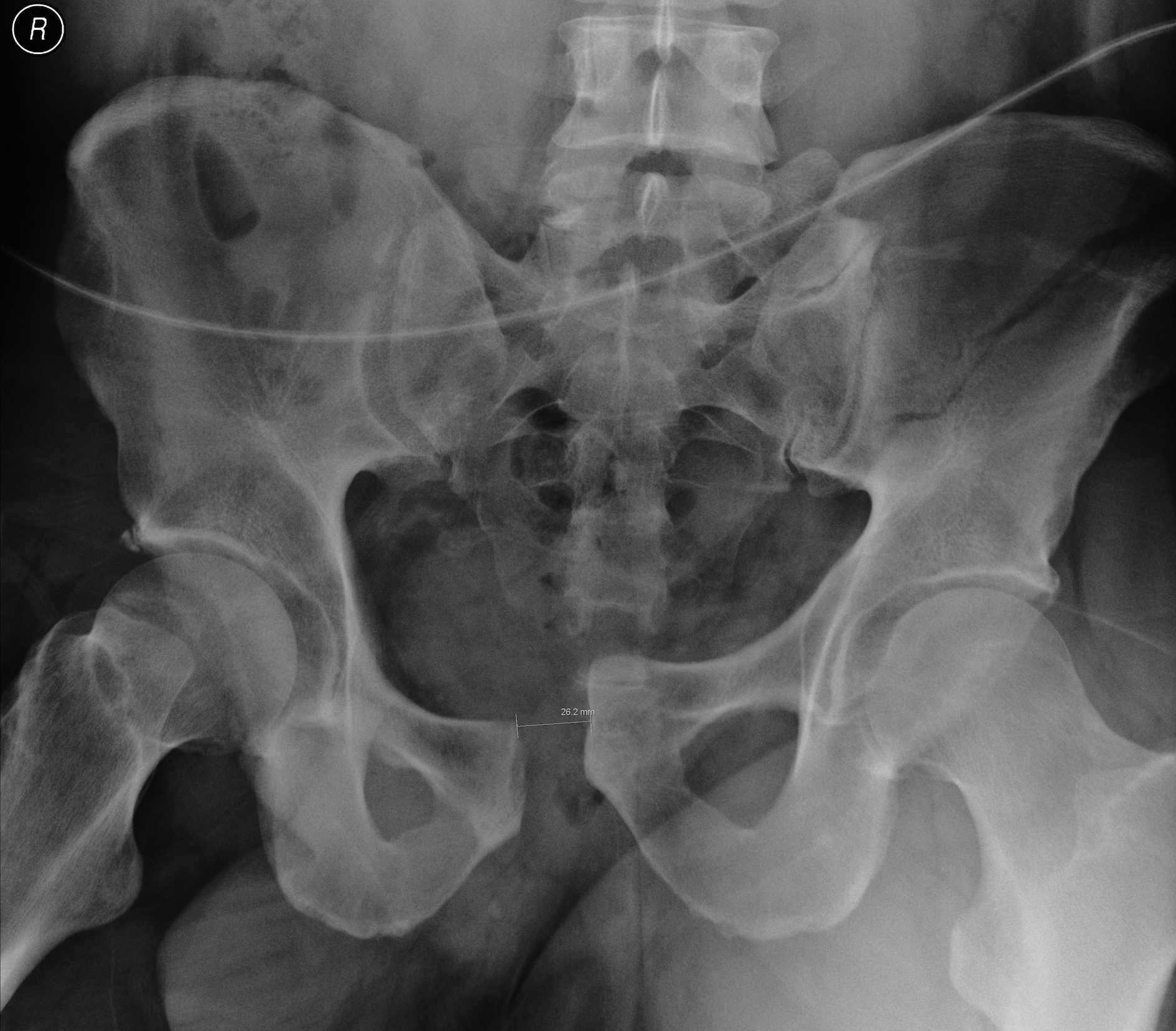
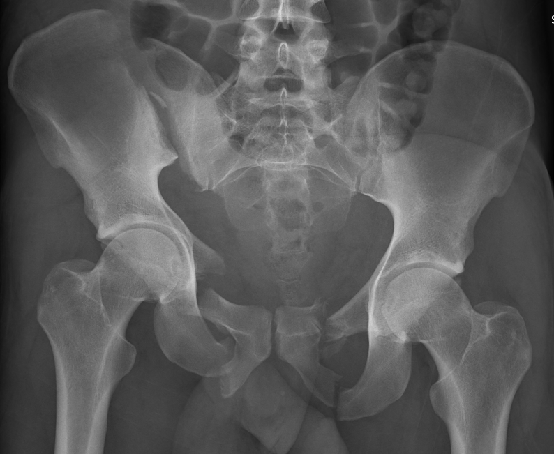
X-rays
Inlet view
- 40o caudal
- shows AP displacement of sacrum and anterior ring
- anterior and posterior sacral borders
Outlet view
- 40o cephalad
- vertical displacement of sacrum relative to ilium
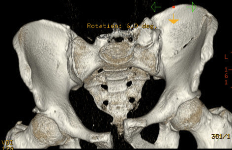
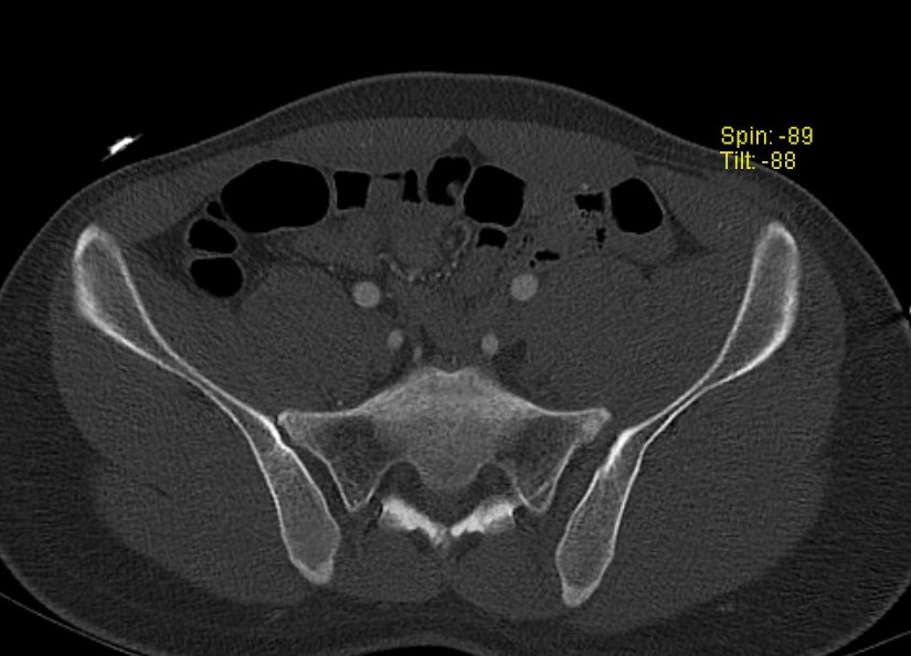
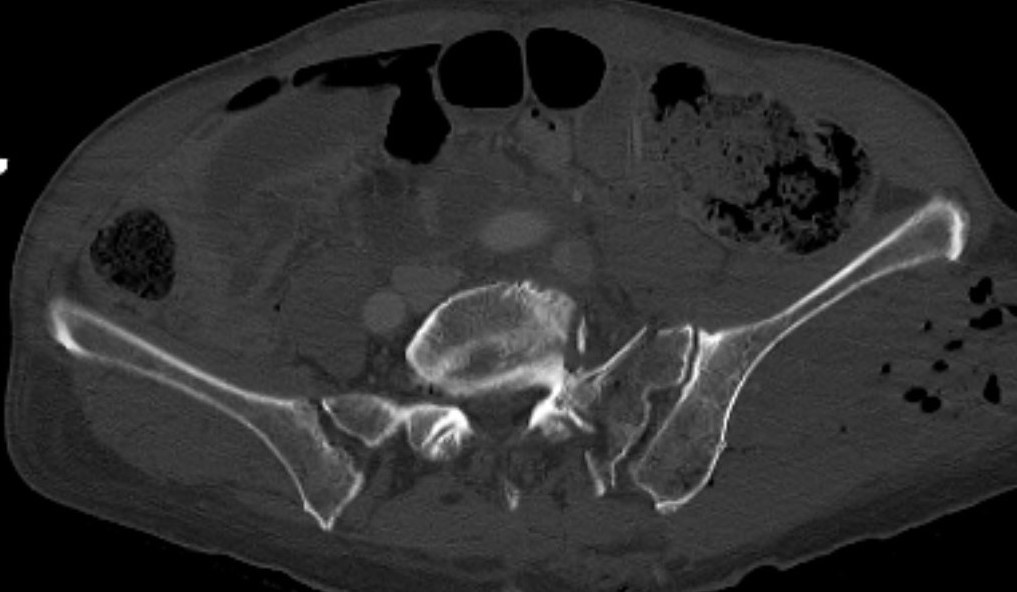
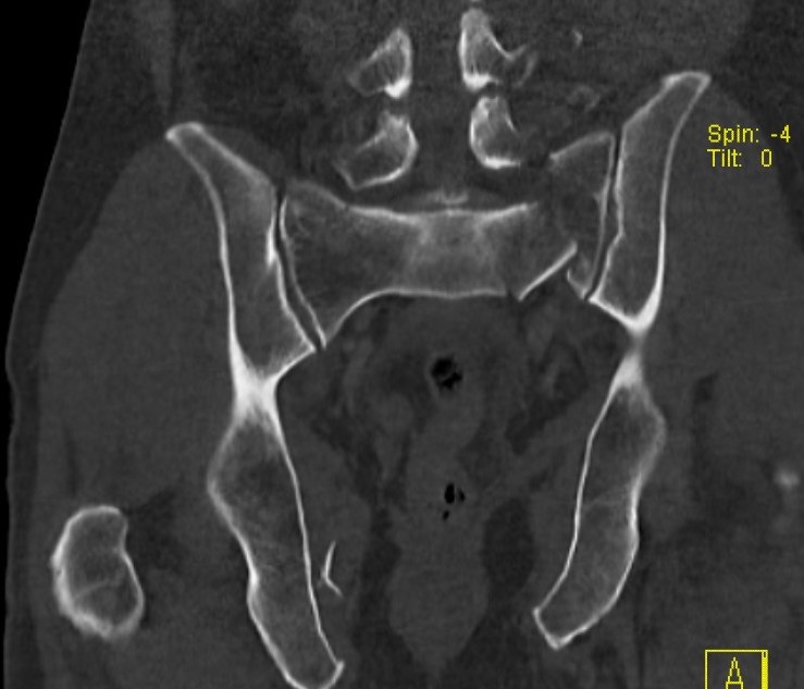
CT scan
Associated injuries
Pelvic vascular injury - arterial / venous
Injuries to urethra / rectum / vagina
Nerve injury
Compound / Morel Lavallee
Pelvic vascular injury
Incidence
- 60% lateral compression
- 52% APC
- 40% vertical shear
Injury pattern
- 15% arterial
- 85% venous
Arterial bleeders
Internal pudendal artery most common
Iliolumbar / SGA / IGA / lateral sacral / internal iliac
Retroperitoneal veins / bone bleeding
85% of bleeding
Visceral injury
Urethra / rectum / vagina / rectum / peroneum
- vaginal and rectal examinations
- looks for blood at urethral meatus
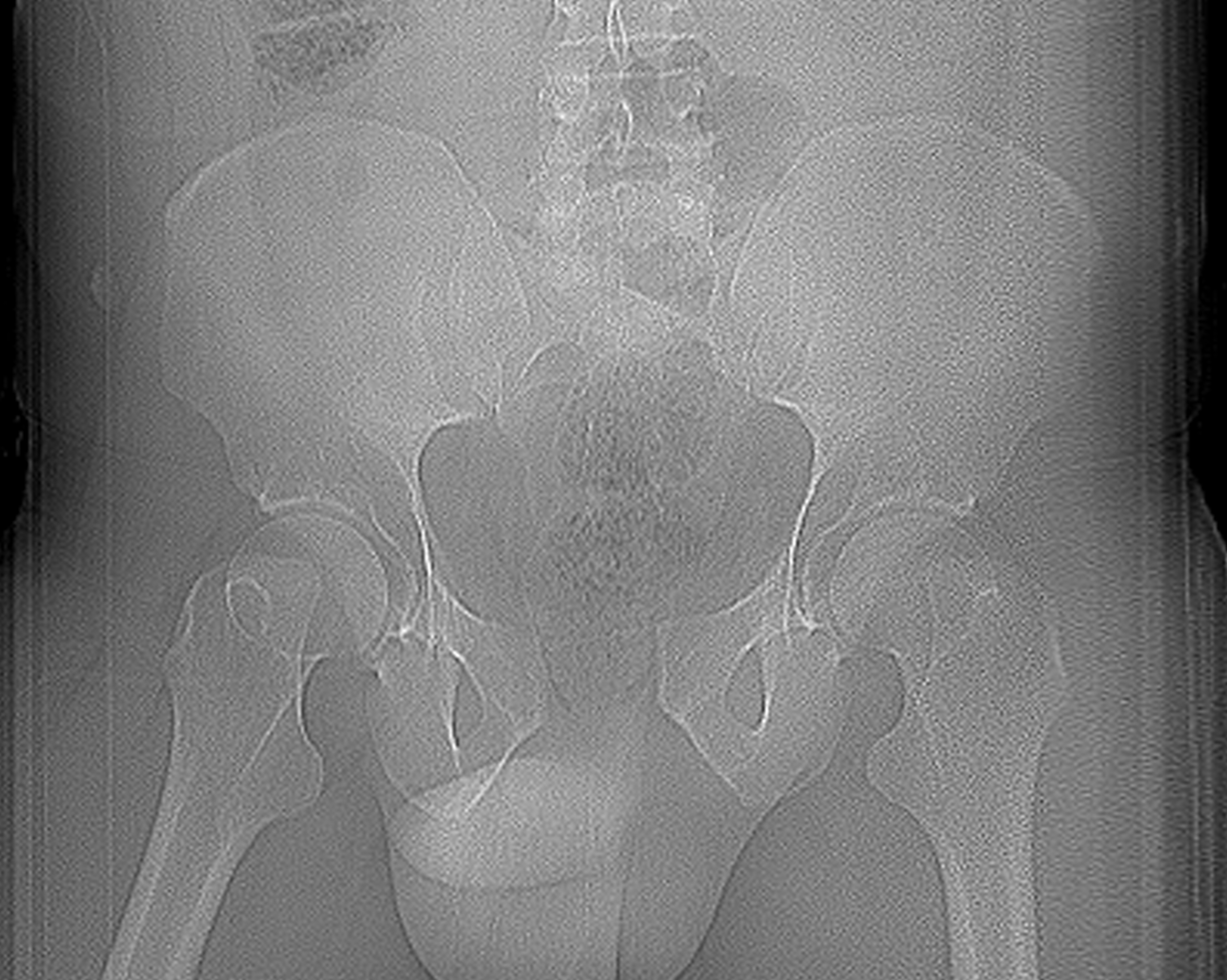
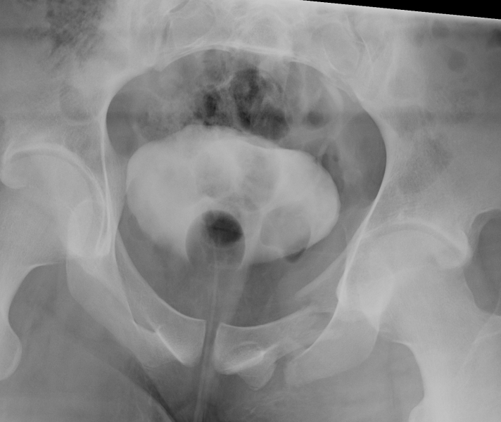
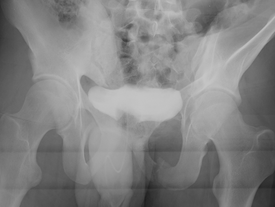
Retrograde urethrogram indicated for blood at meatus +/- retropubic catheter
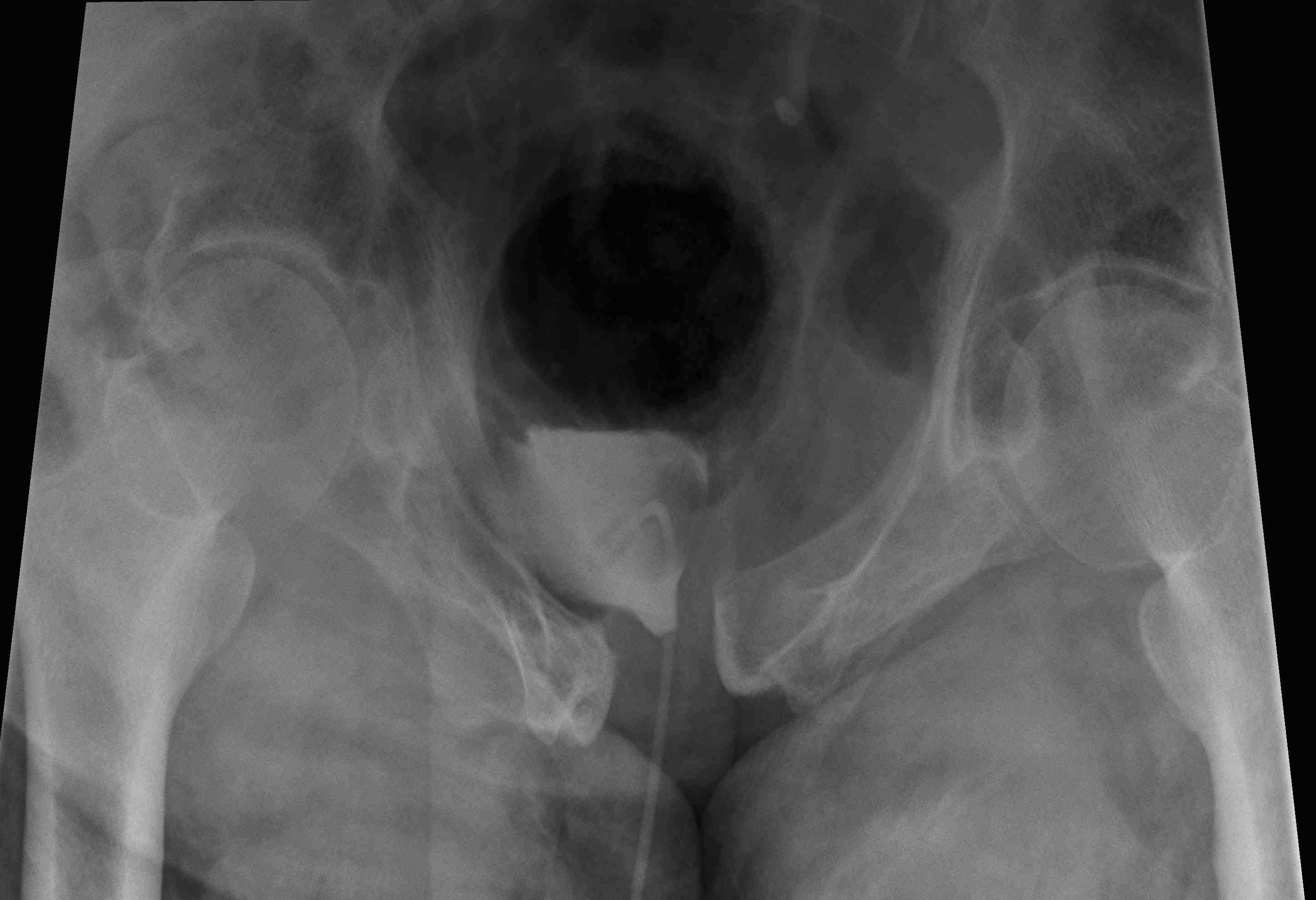
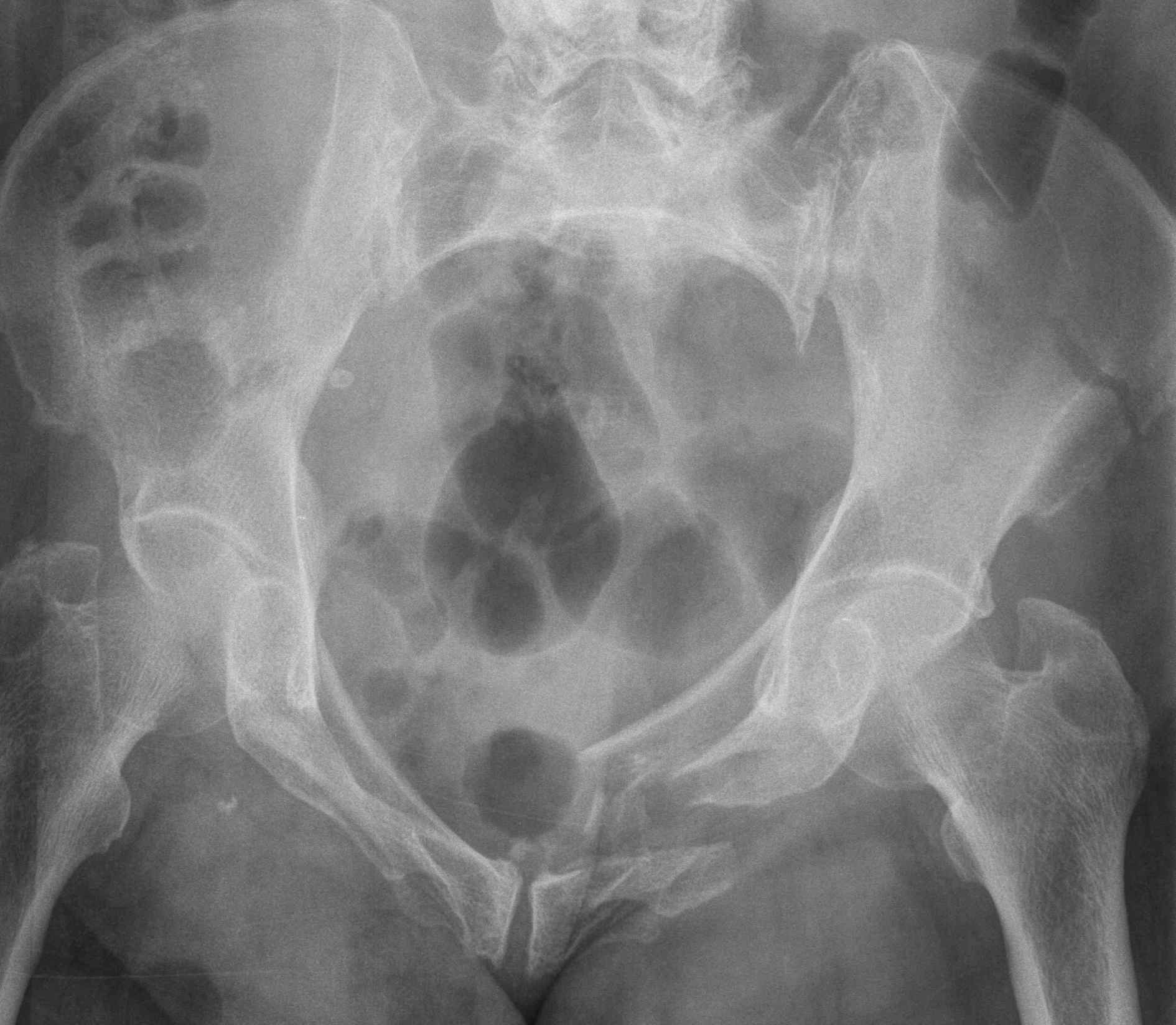
Retrograde urethrogram in setting of APC pelvic fracture
Neurological Damage
Denis classification of sacral fractures
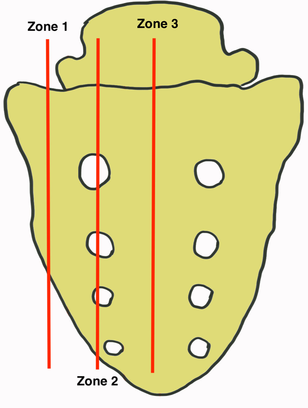
| Site of fracture | Incidence of neurological deficit | Nerve injury | |
| Zone 1 | Lateral to foramen | < 7% | L5 nerve root which is superior to sacral alar |
| Zone 2 | Through foramen | 30% |
S1 / S2 Difficulties with voiding Pudenal nerve numbness |
| Zone 3 | Spinal canal | 60% |
Cauda equina Loss of bladder and bowel function Sexual dysfunction |
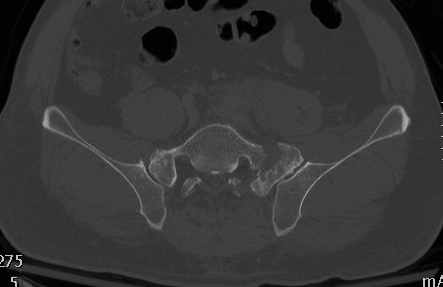
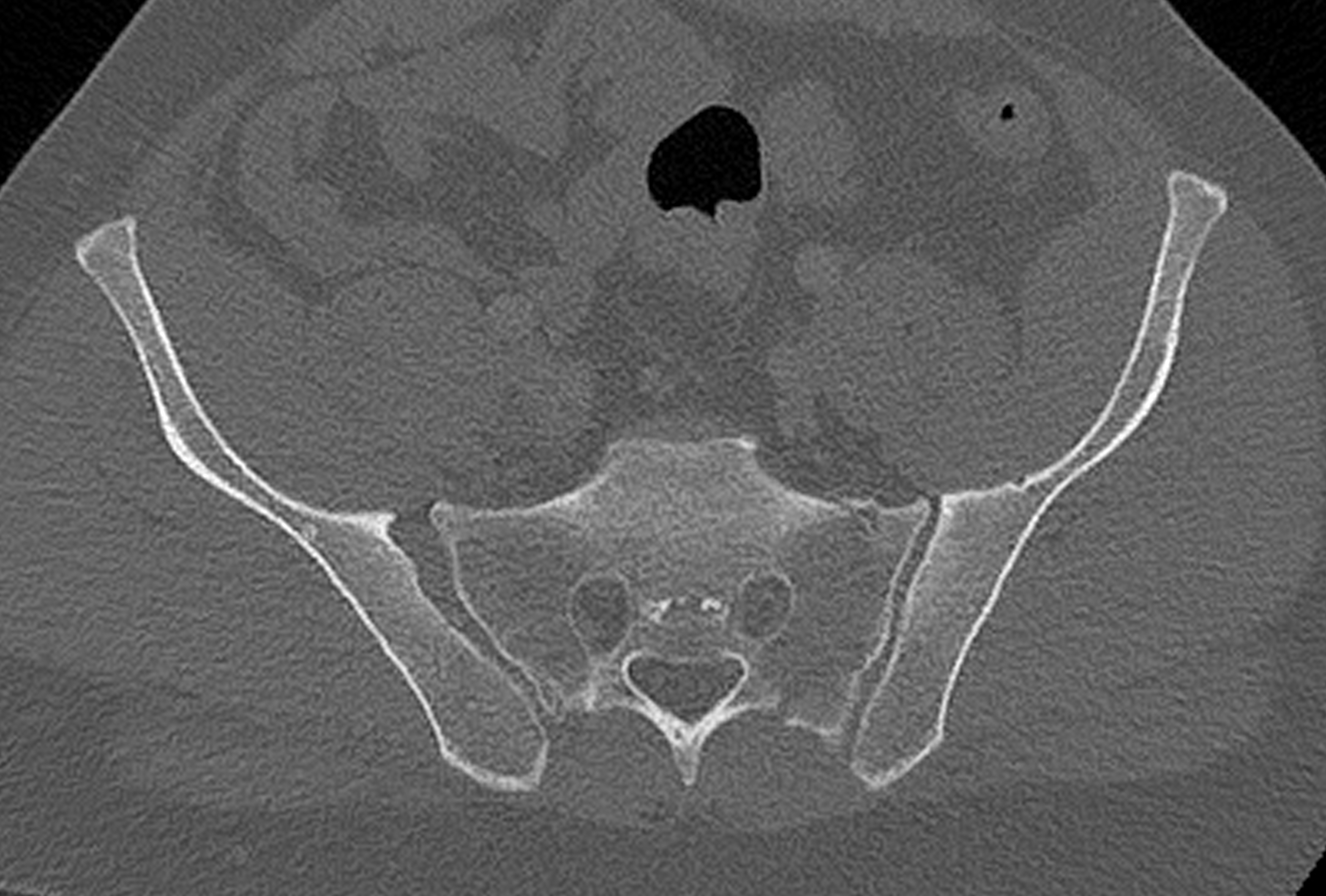
Denis Zone 2 sacral fracture
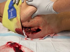
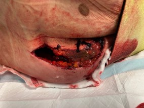
Morel - Lavallee Lesion
Definition
Skin degloving
- predisposes to infection
- found on the thigh in lateral compression fractures
- found in the lumbar area in APC or vertical shear
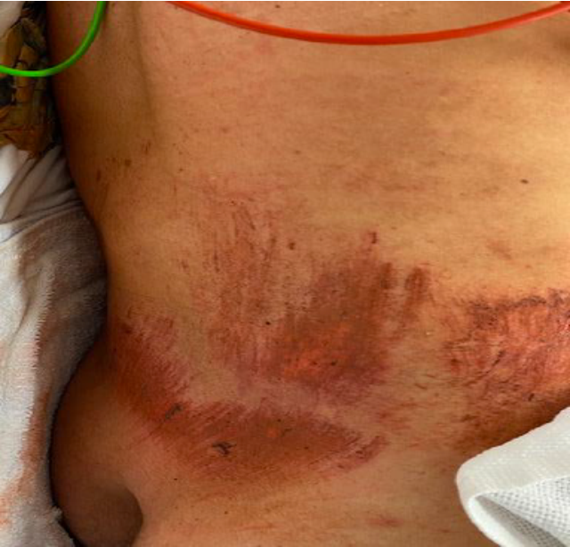
Morel-Lavallee
Beckmann et al Emerg Radiol 2016
- ML lesions seen in 12% of pelvic fractures based on CT
- most common in vertical shear (34%)
- occur in 12% of APC and LC fractures
Compound wounds