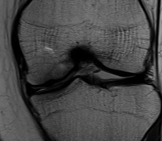
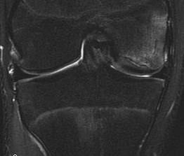
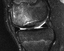
MRI
Plan
Probably unstable
- need to mobilise
- debride base
- bone graft
- fix securely in situe
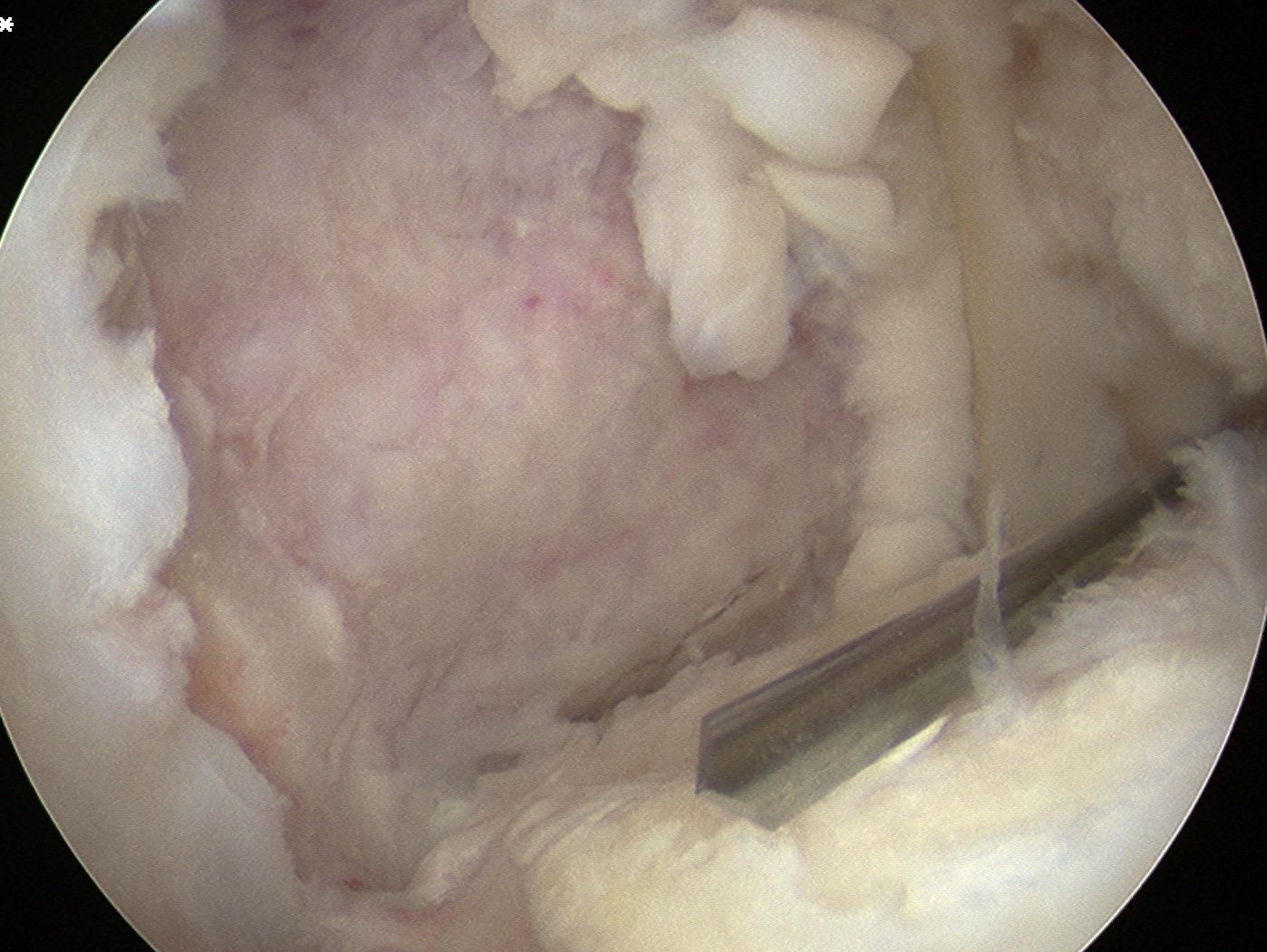
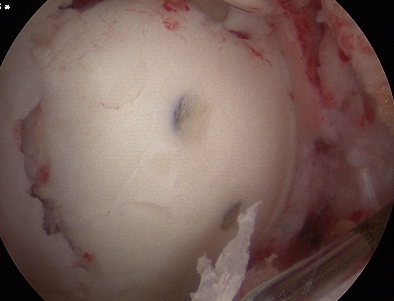
Arthroscopy
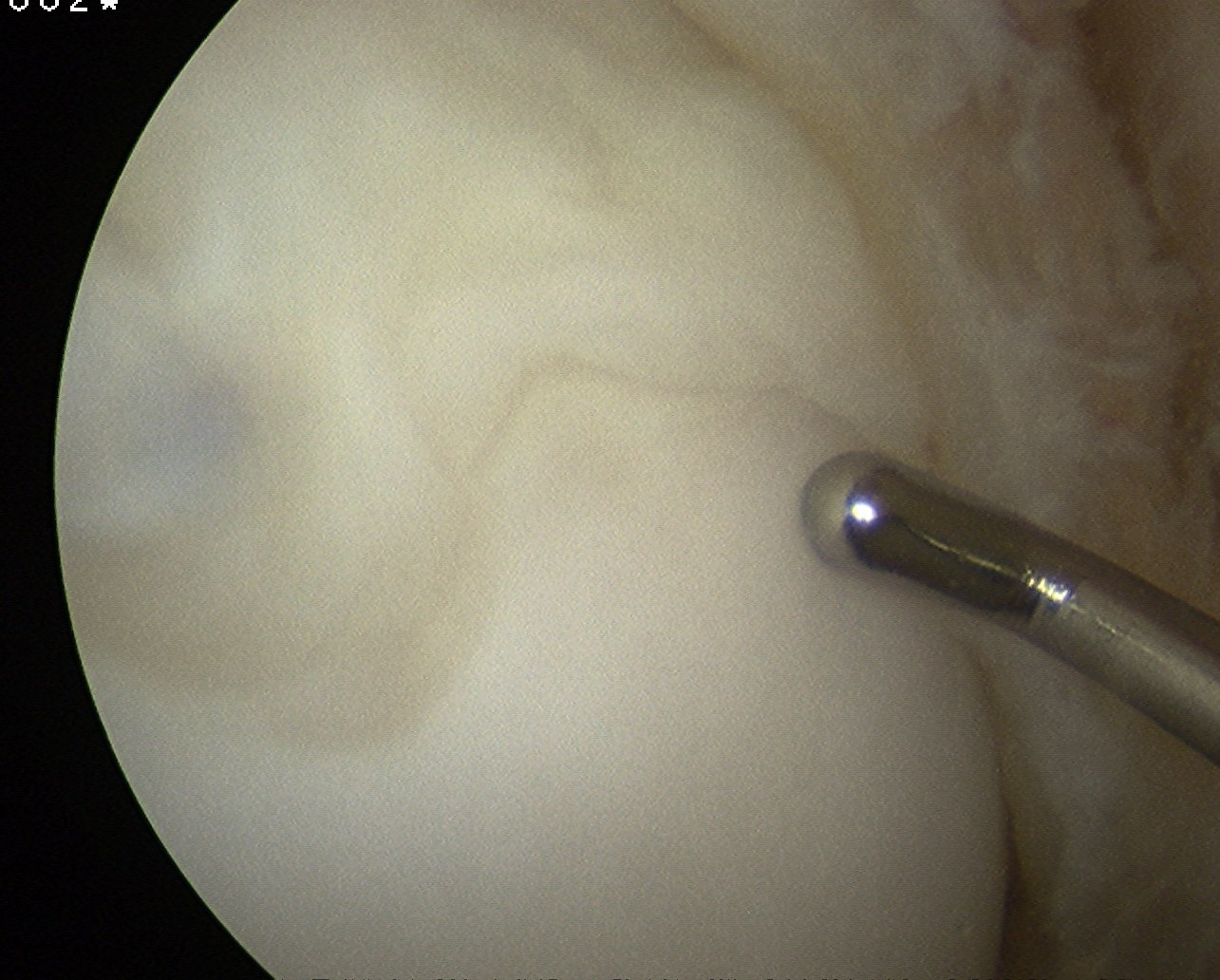
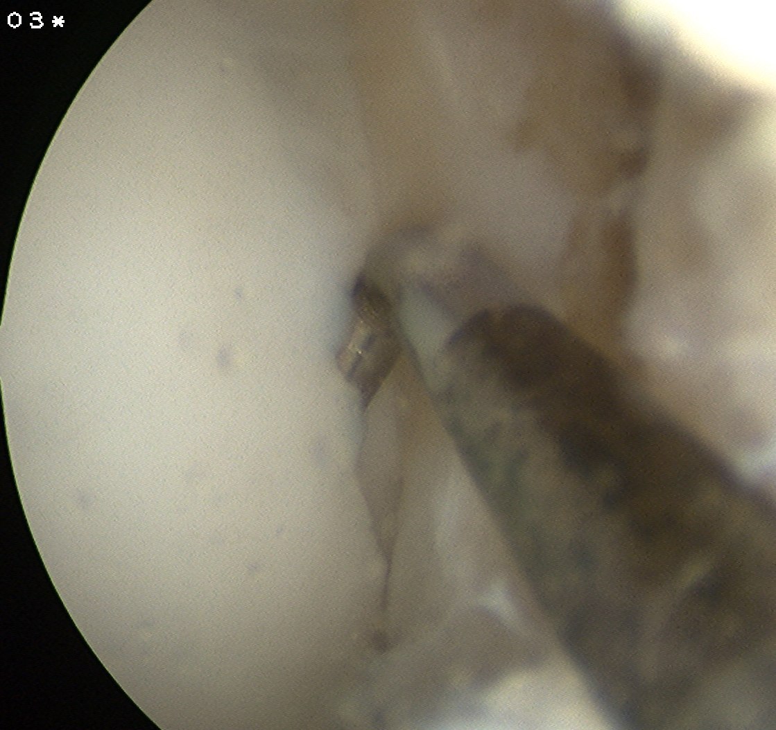
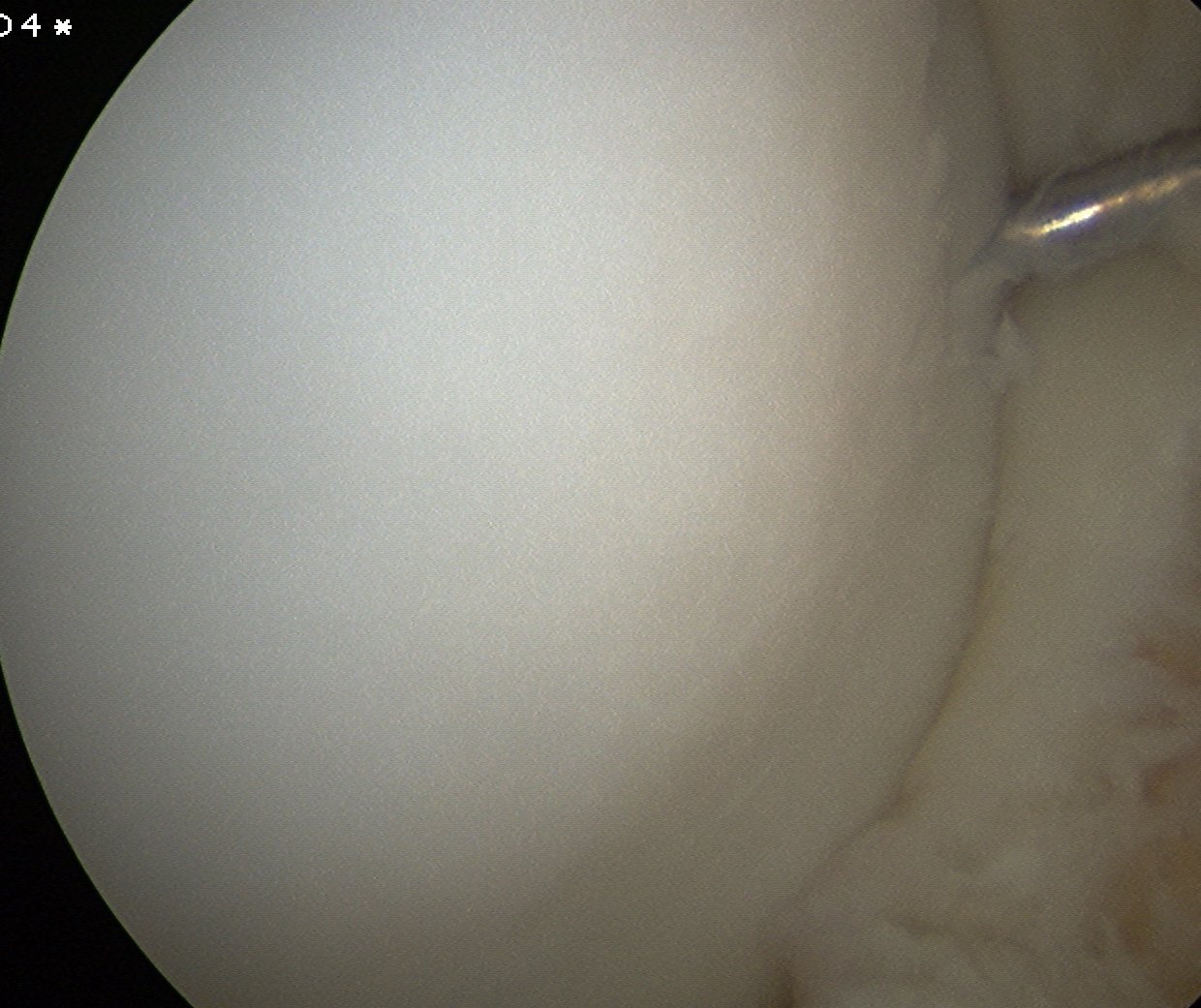
Arthroscope in lateral portal
- instrument through medial portal
- ensure can visualise entire fragment
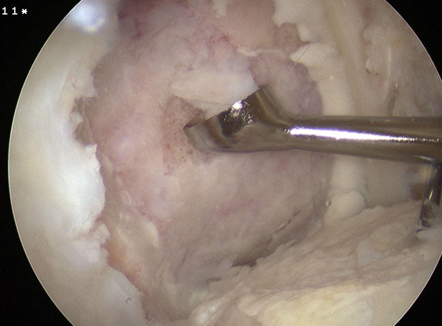
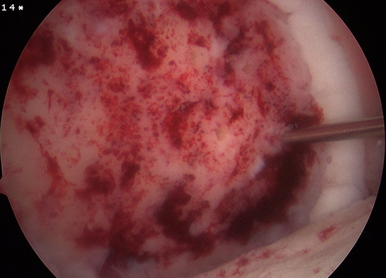
Mobilisation of fragment
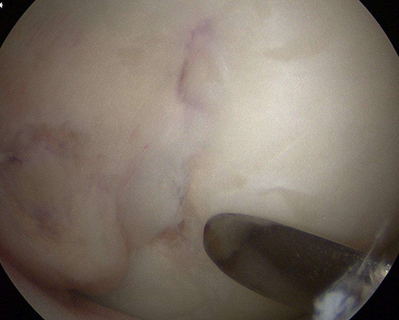
Lesion carefully mobilised with scaple and probe
- left to lever open inferiorly
- want it to stay partially attached
- need to release some fibres of PCL medially
- insert spinal needle from medial knee to hold fragment open
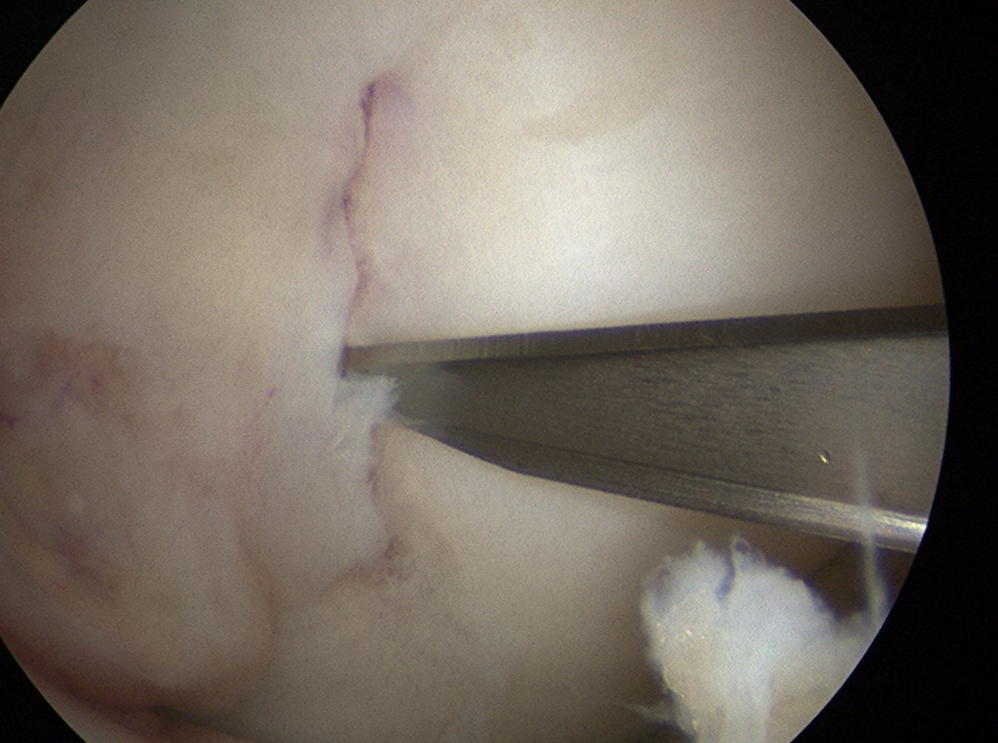
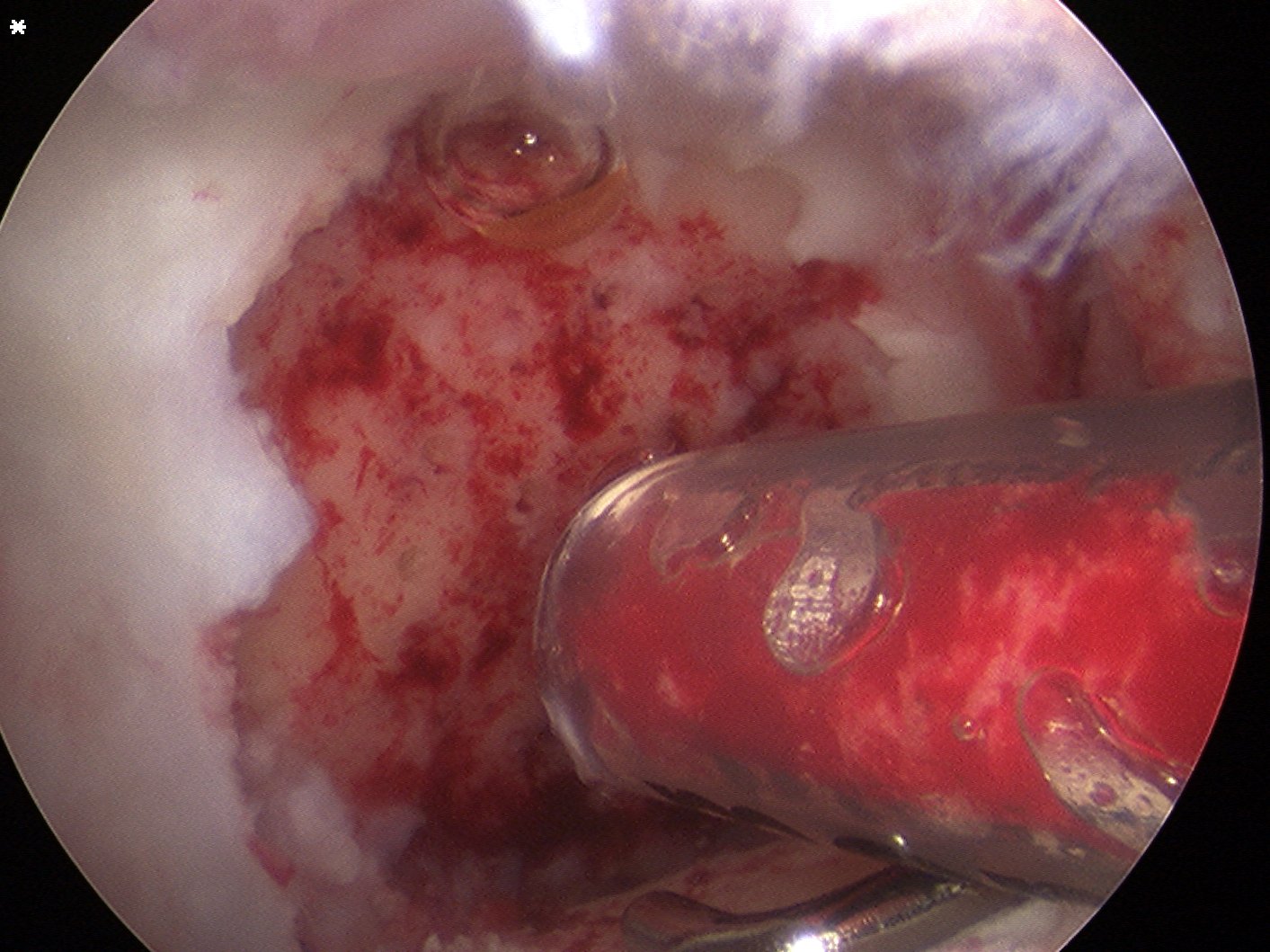
Debride base of lesion
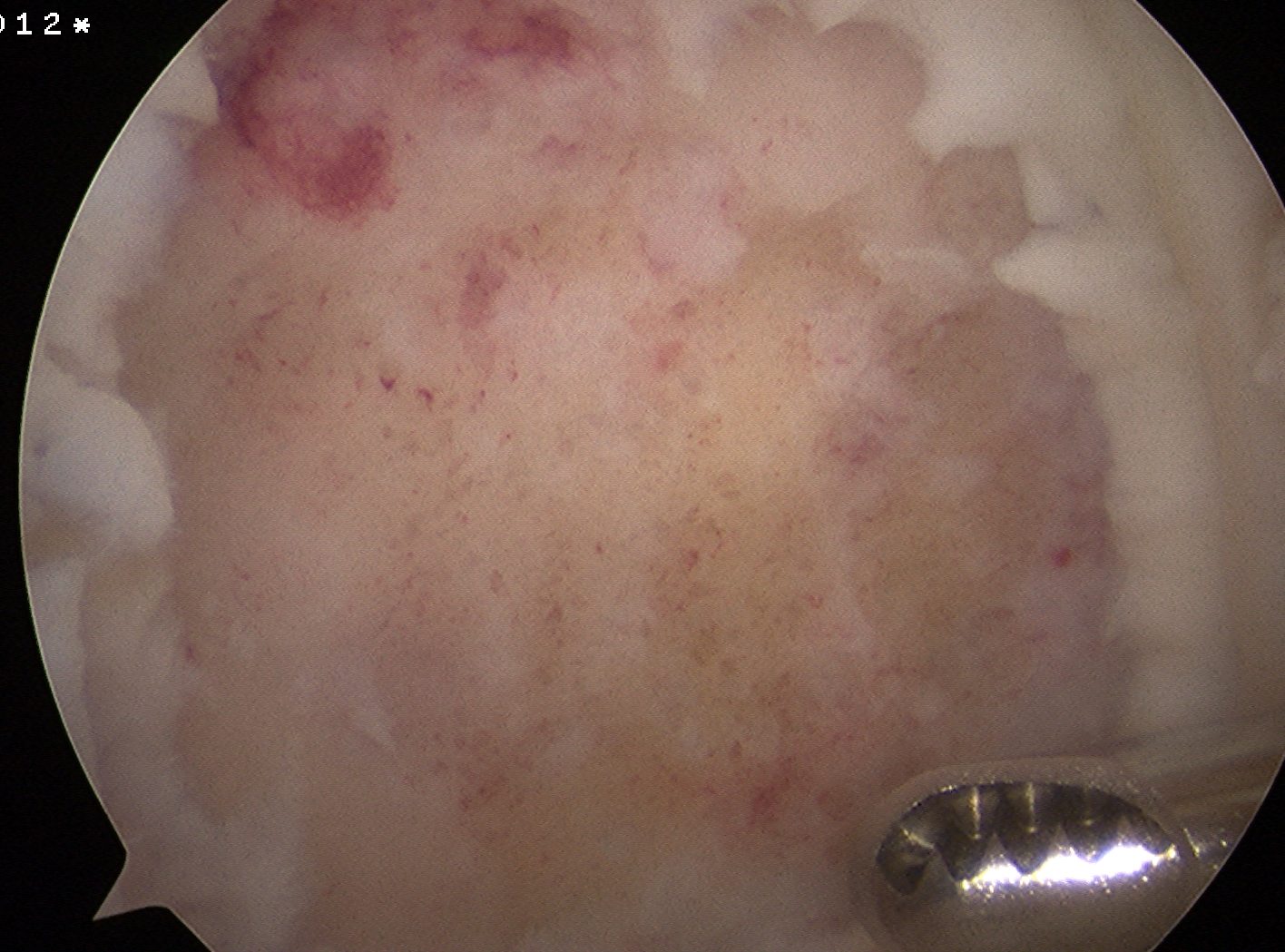
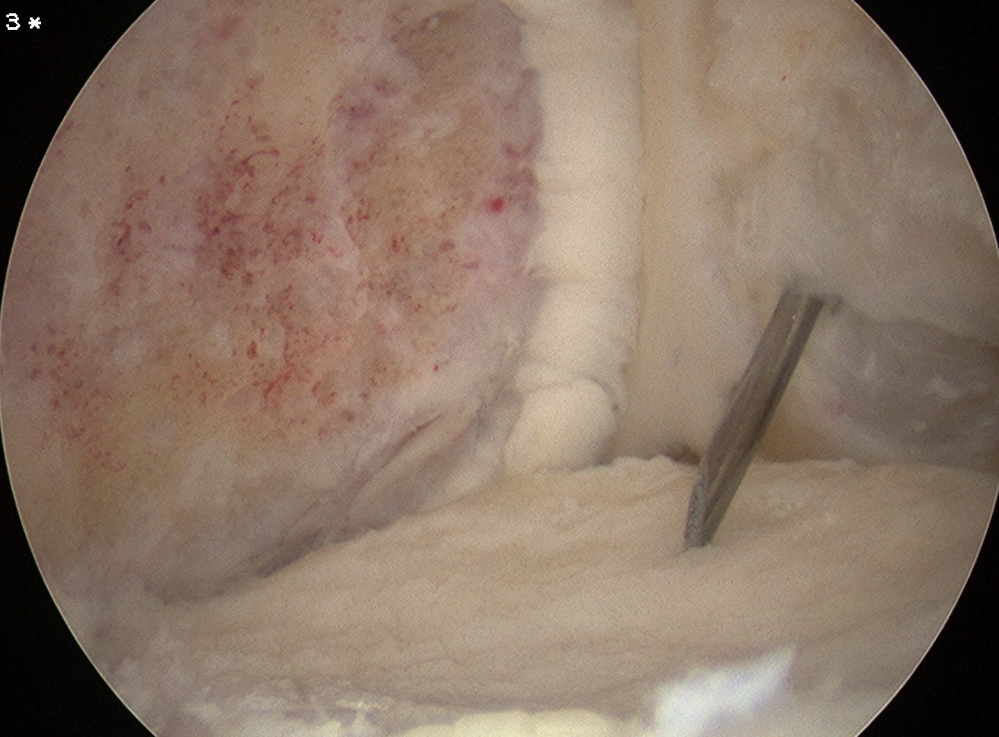
Fibrous tissue removed meticulously from femur with curette and shaver
Take bone graft from medial tibia
- depends on amount of bony defect
- turn into paste / add blood
- put in small syringe that will fit through AM portal
- cut tip off
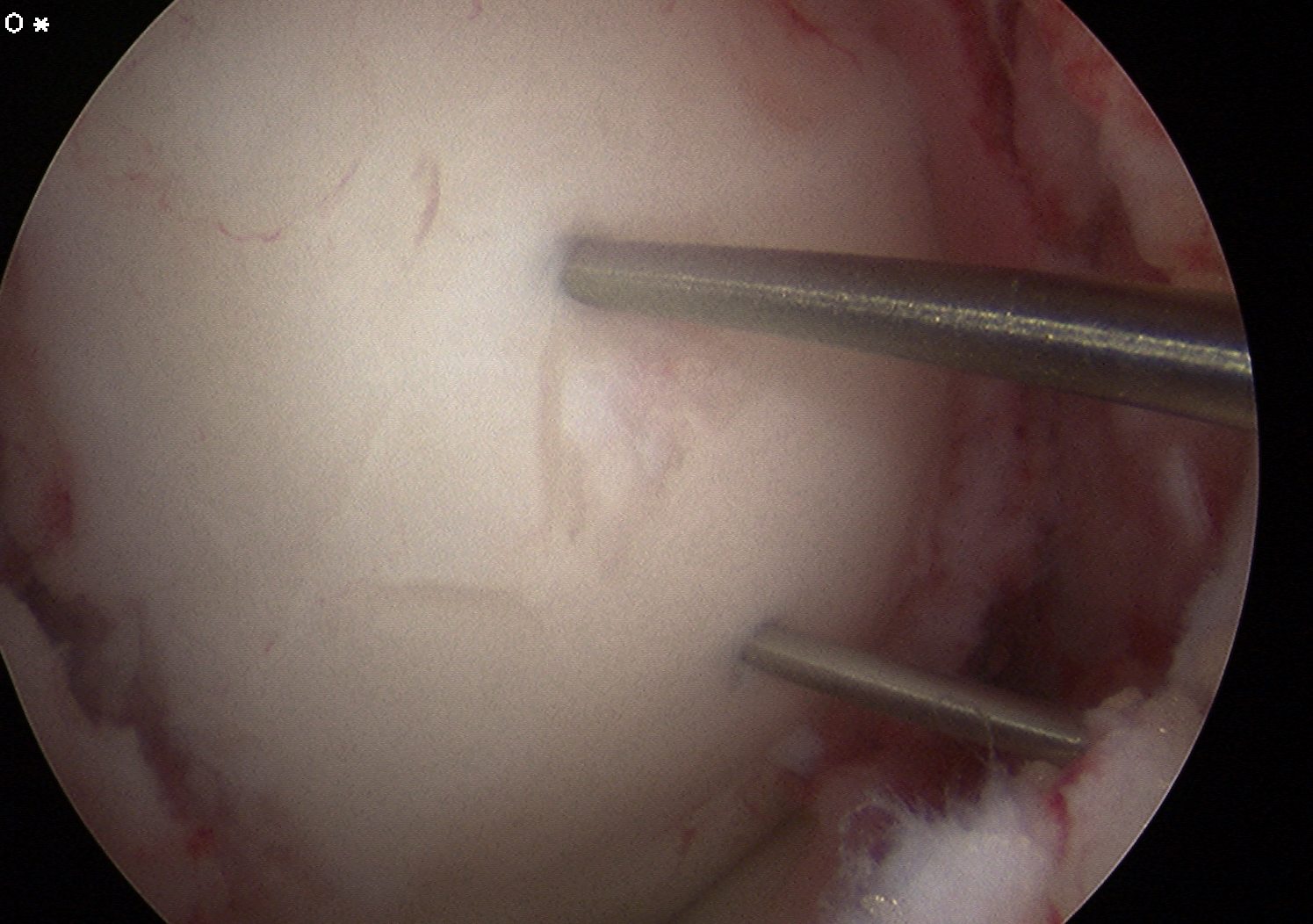
Use K wire to microfracture
- insert bone graft
- immediately reduce fragment
- secure with K wires for cannulated screws
ORIF
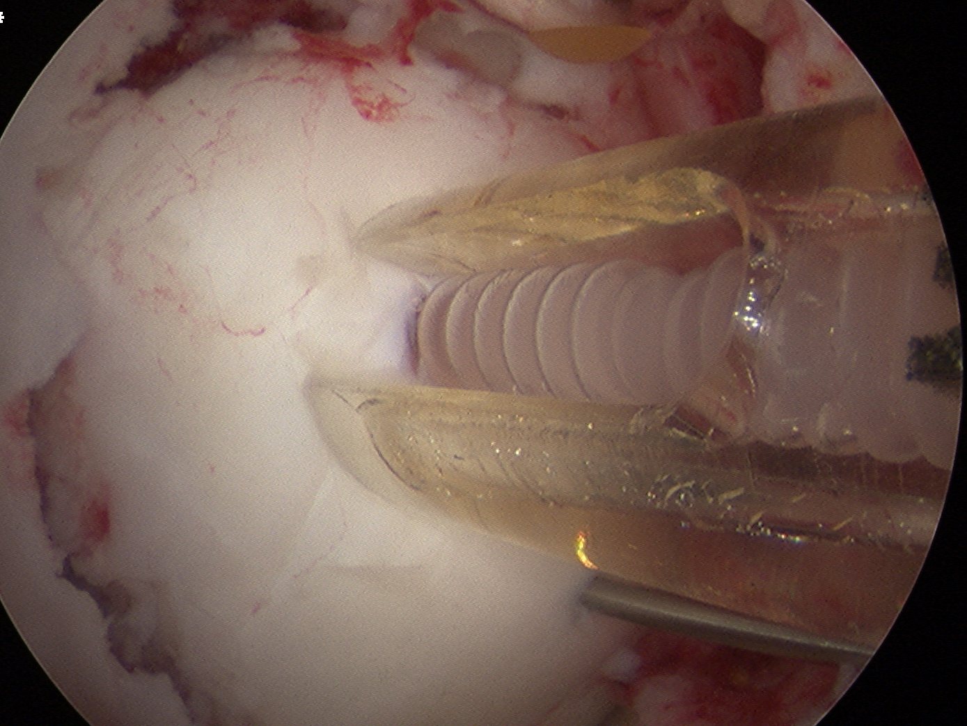
In this case, 2 x Arthrex bioabsorble screws used
- drill and tap over wire
- remove wire
- insert screw and bury head
MRI Follow Up