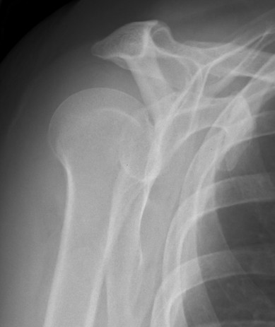
AP Shoulder
Technique
- in plane of thorax
- oblique of GHJ
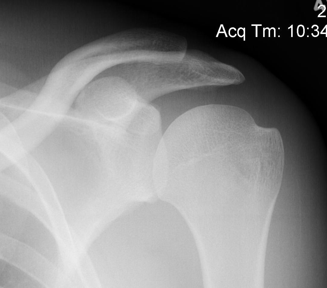
AP in plane of scapula
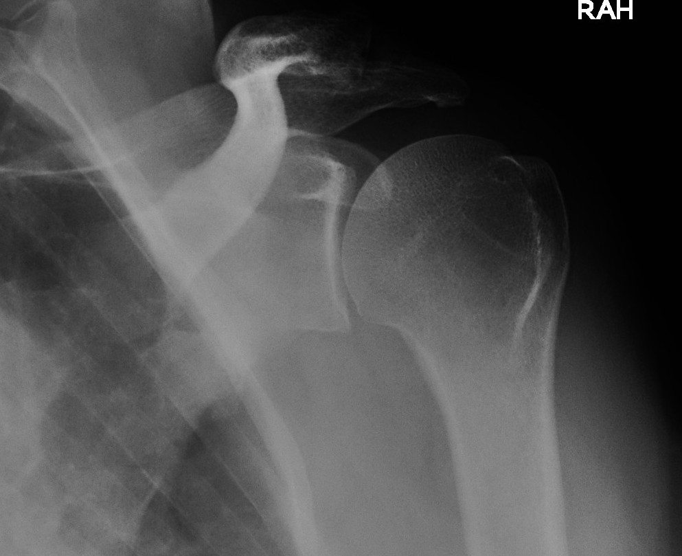
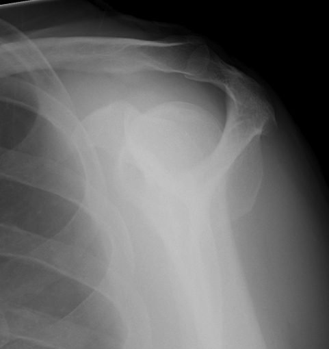
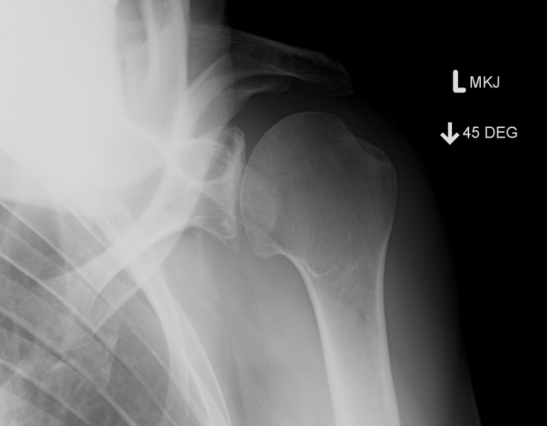
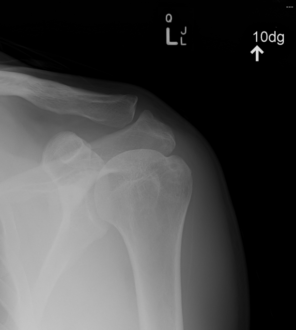
Grashey
- angle 45o lateral
- allows estimation of glenohumeral space
AP IR / ER
Demonstrates Hill Sach's and other humeral head morphology
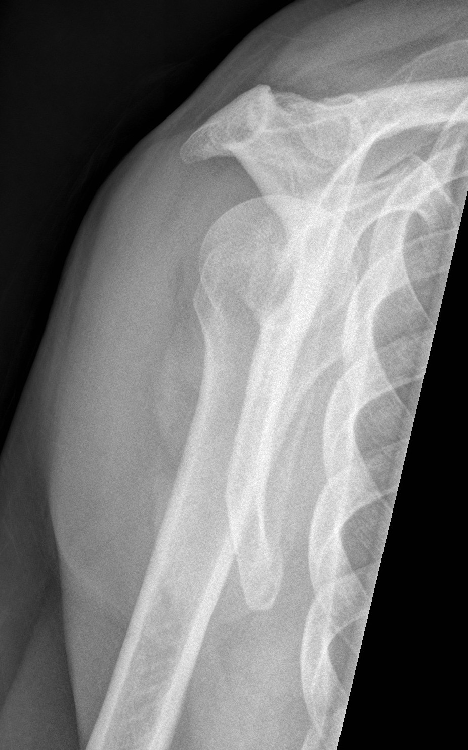
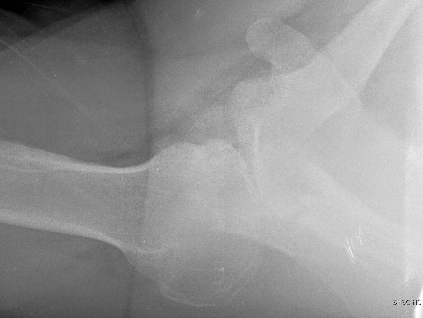
Scapular lateral
Patient erect
- affected shoulder against plate
- rotate other shoulder 45o out of way
- beam aimed along spine of scapula
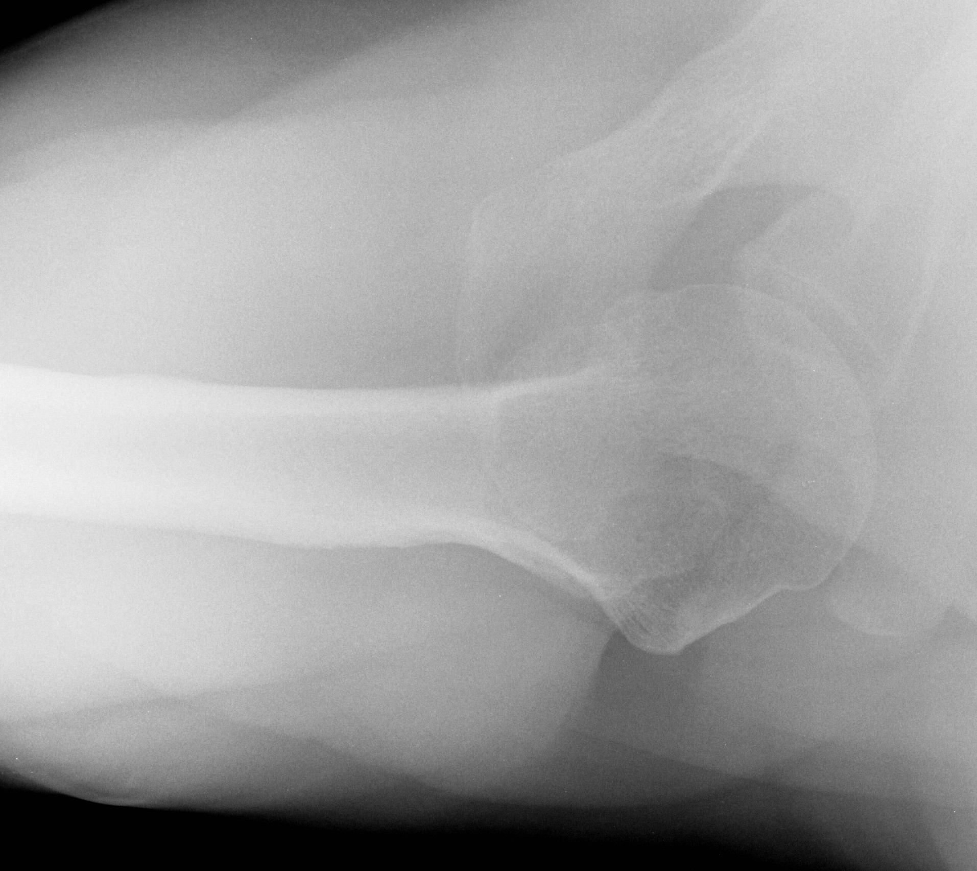
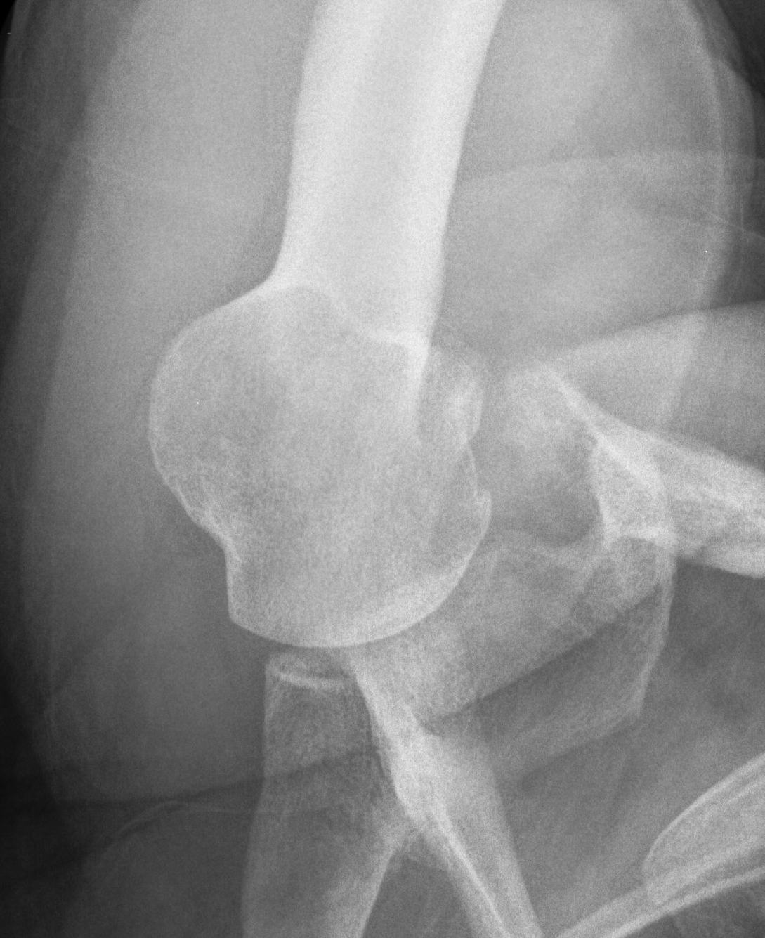
Axillary lateral
Patient seated
- arm abducted
- plate under axilla
- beam angled down towards shoulder
Supraspinatous outlet view
For acromial morphology and impingement
Similar to scapular lateral
- tilt beam caudal 10o
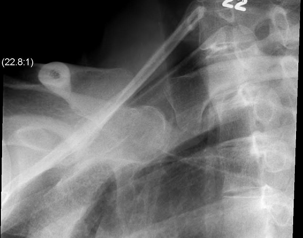
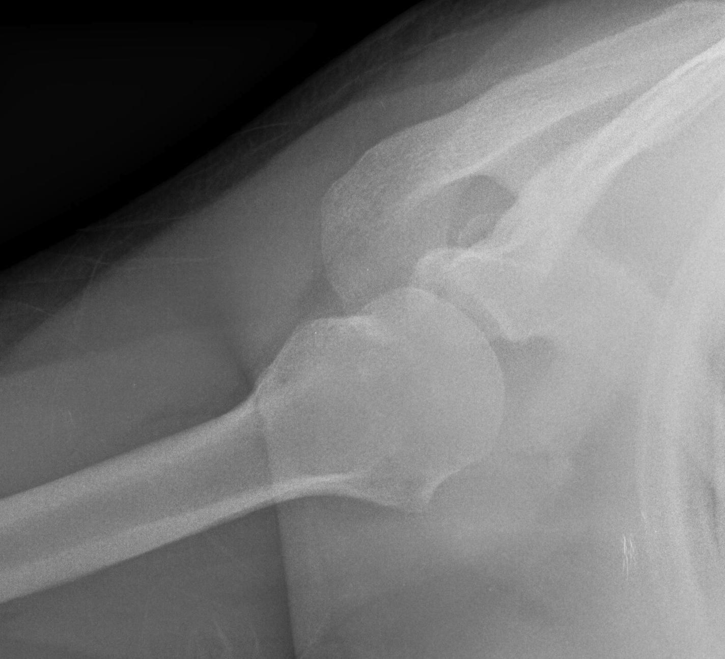
West Point View
Variation axillary lateral
- tangential view anterior / inferior glenoid
- for bony bankart
Patient prone with arm hanging off bed
- plate superior to shoulder
- camera 25° cephalad to horizontal / 25° to long axis body
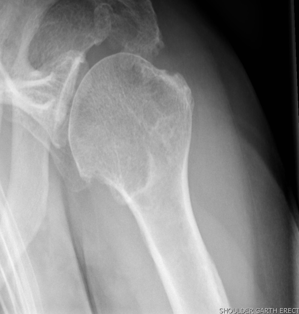
Garth View / Apical Oblique
True AP with 45o caudal tilt
- to show anterior / inferior capsule
- bony bankhart / Hill Sachs
- standing with plate behind joint
- 45° caudal tilt / 45° in coronal
Stryker Notch view
Patient supine with cassette posterior to shoulder
- hand on head, elbow straight up
- beam 10o cephalic aiming at corocoid
Demonstrates Hill-Sach's
Zanca view
ACJ
Patient erect with cassette behind shoulder
- aim beam at ACJ 10 - 15o cephalic
- half strength to not overexpose ACJ
Serendipity view
SCJ
Technique
- prone with cassette under chest
- aim beam 40o cephalic