Definition
Avascular necrosis & subsequent disintegration of lunate
Aetiology
50-75% history of trauma
Occasionally seen in sickle cell / steroid use
Pathogenesis
Vascular Theory
Trauma disrupting vascularity
- single incident with disruption of blood supply
- multiple compression fracture with loss of blood supply to fragments
Linschield
- high incidence of coronal fractures seen on CT
- not apparent on AP film
- disrupts intra-osseous anastomoses
Lunate Vascularity (Gelberman)
Group 1
- 10%
- single incomplete palmar blood vessel
- higher risk AVN
- severe hyperextension may disrupt it
Group 2
- 90%
- dorsal & palmar blood vessel
- well vascularized
- need intra & extraosseous disruption
- low risk AVN
AVN not seen in lunate dislocation
- flap of volar capsule usually remains attached
Mechanical Theory
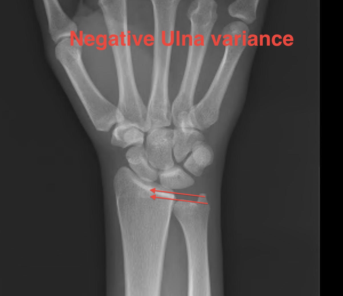
Normal ulna plus
- +2 to -6 mm
- +2 SD mean
Ulna minus variance
- subjects lunate to greater compression & shear forces
- increased radioulnar forces
- seen in 75% Kienbock's
- only 25% normal population
Lichtmann Classification
Stage 1
- no radiological change
- diagnosed on bone scan / MRI
Stage 2
- sclerosis
Stage 3
- collapse / fragmentation
A: Normal carpal height
B: Loss of carpal height / scaphoid flexed / capitate migrates proximally
Stage 4
- degeneration
- pan carpal arthritis (radiocarpal / midcarpal)
Epidemiology
Occurs in young active adults
- age 20-40
- usually dominant hand
- rarely bilateral
- men > women
History
Gradual onset of stiffness & pain
50% history of trauma
Examination
Decreased ROM
Poor grip strength
Tender over lunate
Passive dorsiflexion middle finger gives pain
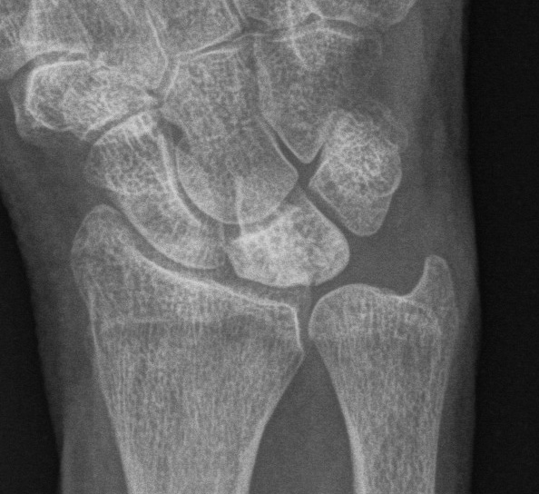
X-ray
Kienbock's
- progressive changes of AVN
- mottling / collapse / OA
- look for scaphoid flexion / capitate descent
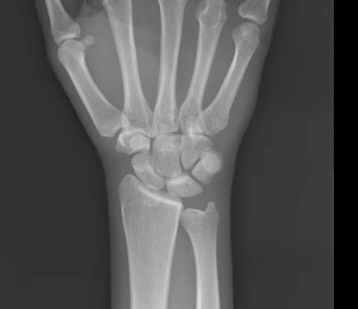
Ulna Variance
Supination and pronation alter values
- need zero rotation view
90 / 90 view
- PA film with wrist in neutral
- elbow 90° / shoulder Abducted 90°
Line from lunate fossa and ulna head
- mean ulna variance is 1 mm (range 2 - 4)
Bone Scan / MRI
Demonstrate AVN
CT
Often show coronal fracture with palmar fragment extruded
- ? Cause of decreased palmar flexion
NHx
Usually one of progressive collapse
- cccasionally can arrest and even reverse
- these patients may not be seen
- the usual patient presents late
- makes interpreting treatment options difficult
Non-operative Management
Splint
Rarely effective
- Stage 1
- trial of immobilisation for 3/12 to aid revascularisation
1. Stage II / IIIA Negative Ulna Variance
Radial Shortening ~ 2mm
Theory
With negative variance often have thick healthy TFCC
- can tolerate loading well
Radius normally takes 80% of load
- with ulnar minus is increased to 96%
Redistribute stresses
- 2mm: 20% decrease radiolunate load
- 4mm: 40% decrease in radiolunate load
- less stress on lunate / revascularisation
- relative lengthening of tendons may decrease compressive forces
Aim
- aimed for neutral or +1 mm ulnar variance
- if >1mm positive then risked ulnar abutment
Technique
- volar approach
- resection of desired amount
- can use cutting guides which give 2 parallel oblique osteotomies of set distance
- ensure not violating DRUJ
- application volar plate
- can be done dorsally but plate can become problem
Results
Quenzer J Hand Surg 1997
- 68 patients
- diminished pain 90%
- increased grip strength 75%
- increased ROM 50%
- 1/3 had signs lunate revascularisation
2. Stage II / IIIA with Neutral or Positive Ulna Variance
Capitate Shortening + Capitohamate fusion
Aim
- unload the lunate
Result
Almquist Hand Lin 1993
- 83% revascularisation and healing
Vascularised Bone graft
Indications
- best success in stage II / precollapse
- can combine with capitate shortening
Options
- 2nd dorsal intermetacarpal A & V
- distal radius pronator quadratus pedicle
- dorsal distal radius on pedicle
3. Stage IIIB
Limited fusion
A. STT
Theory
- lunate collapsing
- scaphoid takes more of load and goes into flexed position much like DISI
- STT fusion gives stable radial column for load bearing
- prevents radiocarpal degeneration
Technique
- dorsal approach 3/4
- take scaphoid out of flexion / extend
- K wire into position
- fuse to trapezium and trapezoid using bone graft from distal radius
B. 4 corner fusion
Theory
- provides ulnar load bearing column
Problem
- lunate is poor quality / necrotic
Proximal Row Carpectomy
Always consider adding denervation
4. Stage IV
PRC
- contraindicated with severe capitate degeneration
Arthrodesis
- manual worker