Indications
Deformity correction
Technique
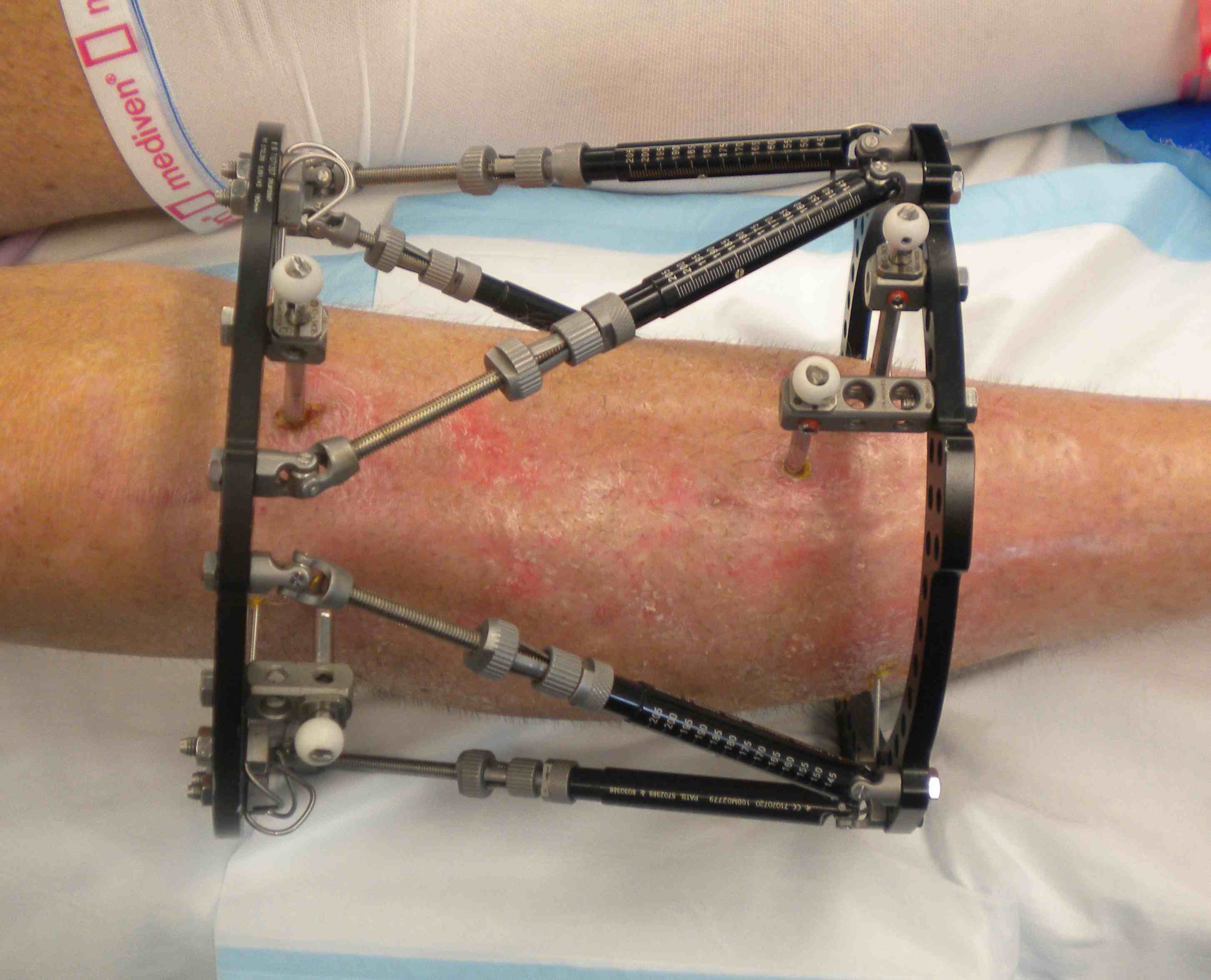
Apply proximal and distal rings
- choose size
- 2 cm from skin all around
- leg will often swell
- most often compression is posterior
- can use an open ring proximally to aid knee flexion
Apply proximal rings
- Centre / Master Tab anterior to tibial crest
- important for calculations
Apply Distal ring
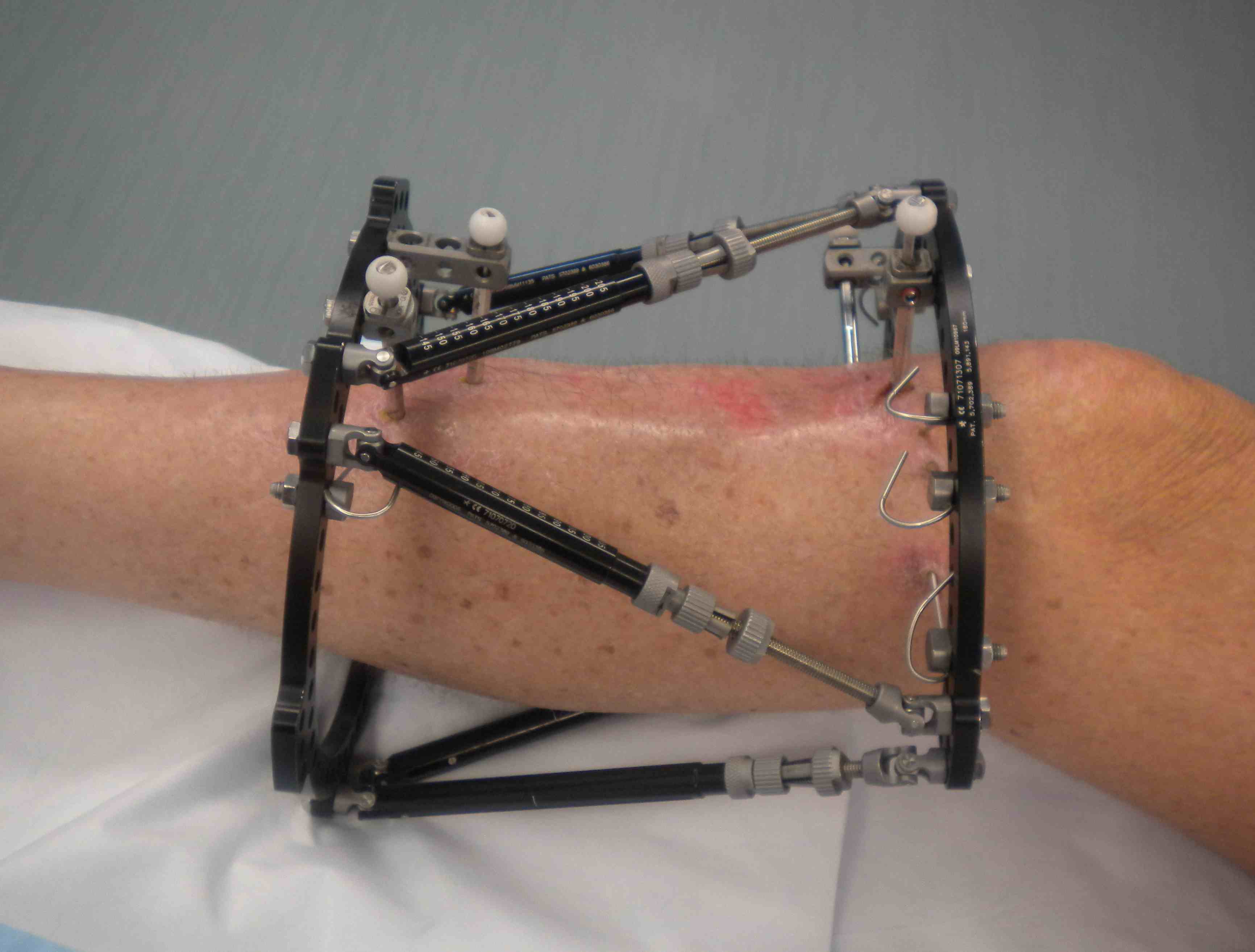
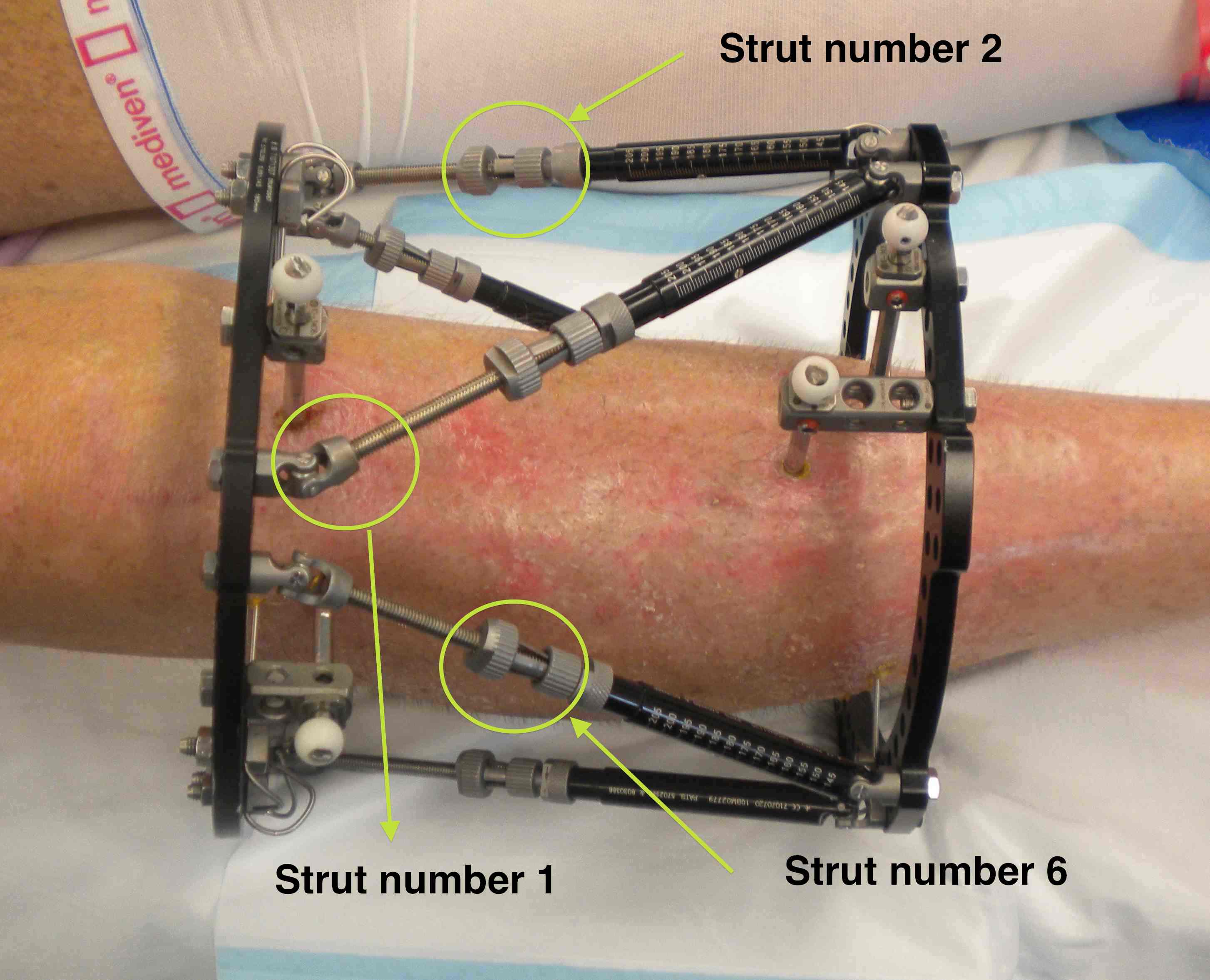
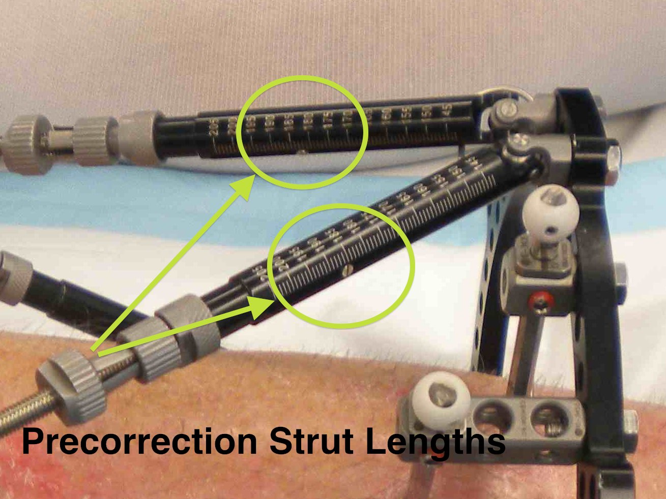
6 x adjustable struts
- come in 3 lengths (short / medium / long)
- need to ensure that are long enough for any correction
- otherwise will have to adjust strut later
- numbered 1 - 6
- no 1 always to right of proximal strut
Intra-operative Correction
- can perform
- new computer program
- struts can be of different lenths
- i.e. the rings do not have to start perpendicular to shaft
Record numbers of each strut
- important for computer calculation
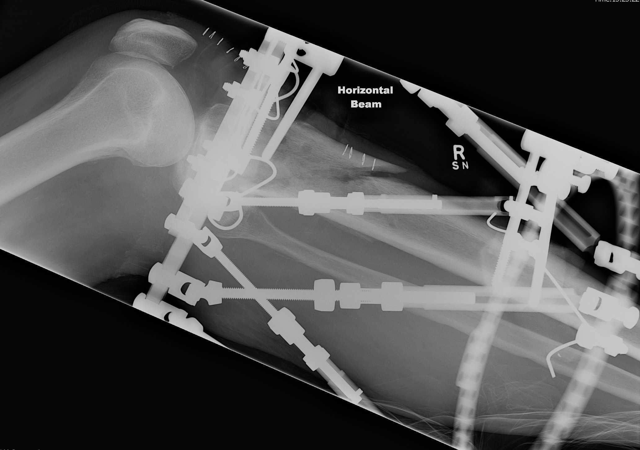
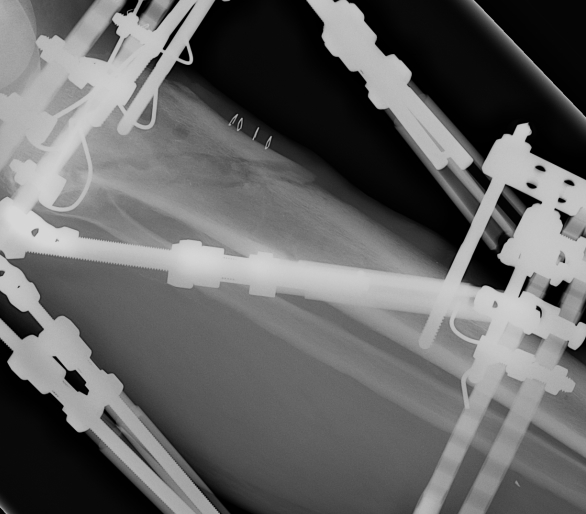
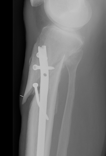
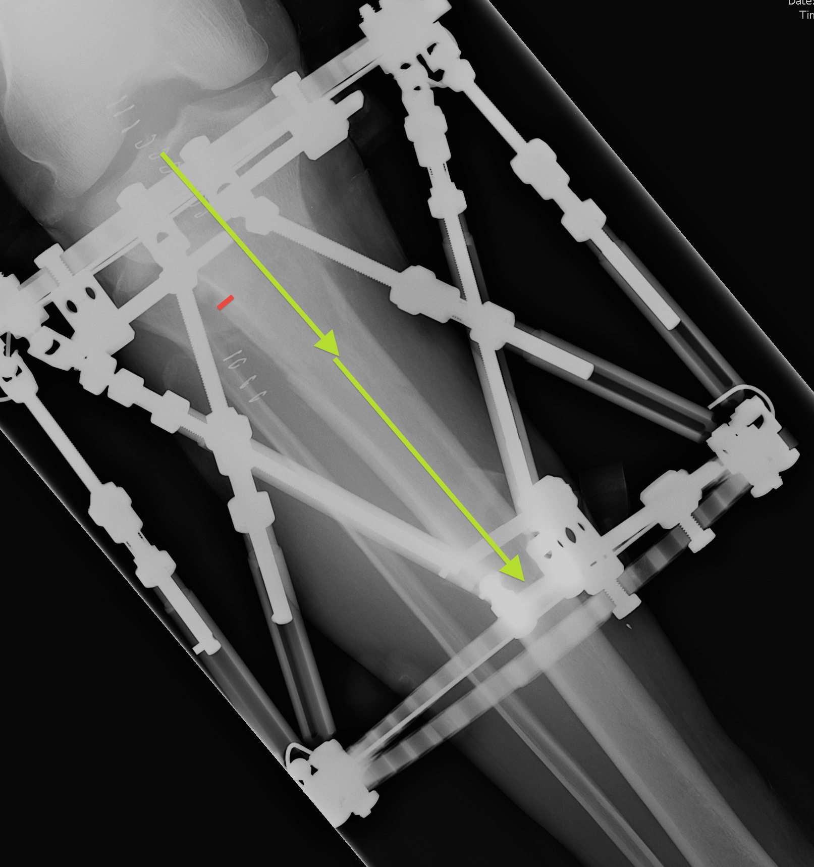
Post operative Xray Measurements
Xray of entire tibia and rings
- xray centred on proximal tab of proximal ring
- AP and Lateral all in one film
- the entire diameter of the rings must be on the film
Set reference fragment
Proximal tibia
- usually the proximal fragment is reference fragment
- distal fragment is moving fragment
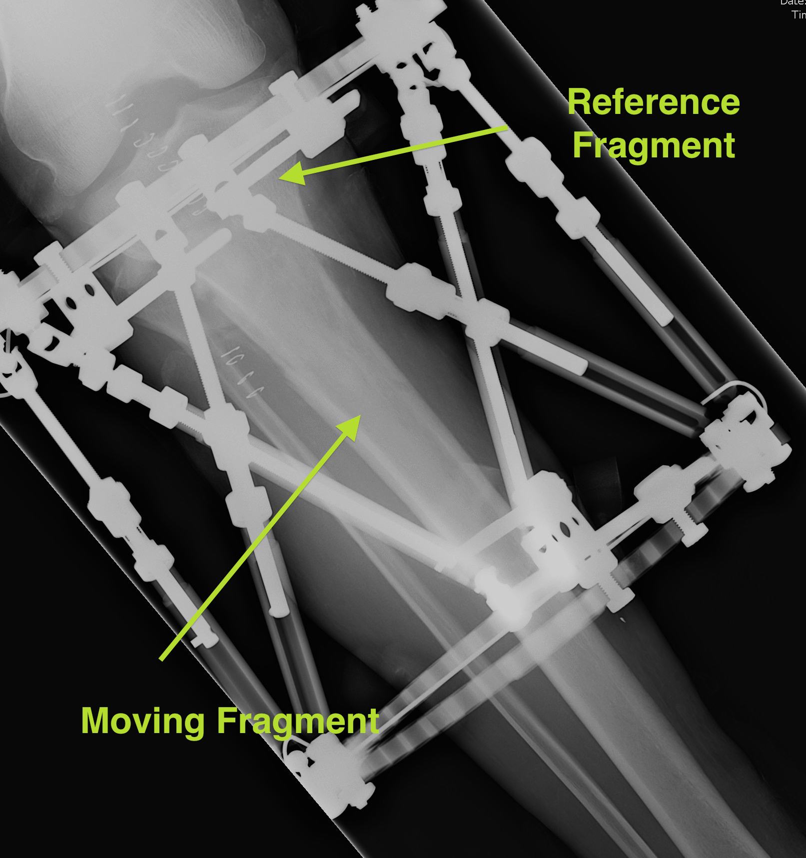
Opposite for Distal Tibial Fractures
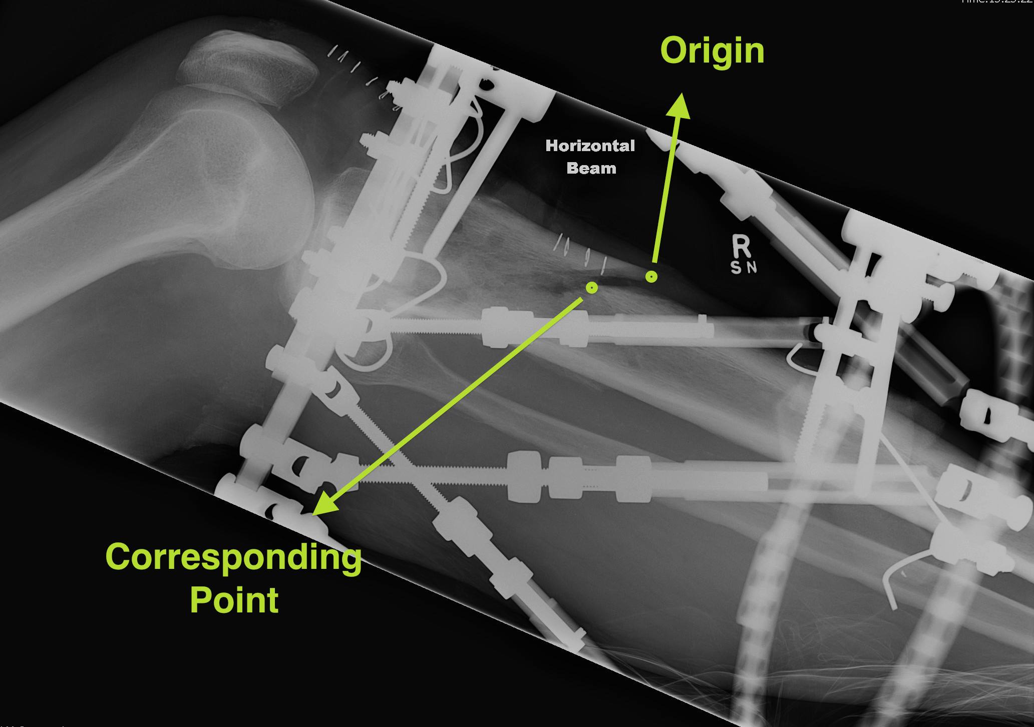
Set Origin / Corresponding Point on AP and Lateral
Origin
- reference fragment
Corresponding point
- on moving fragment
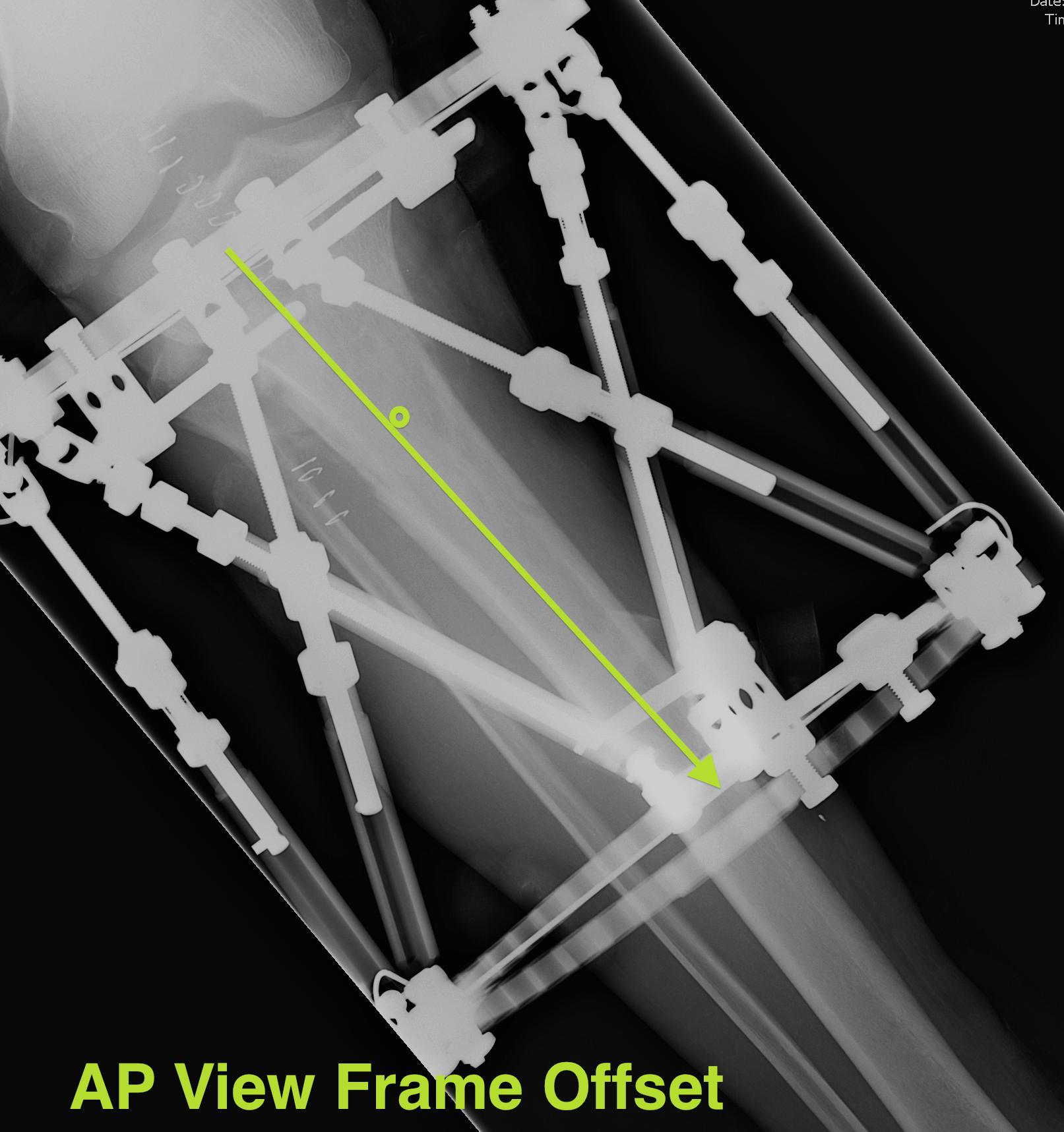
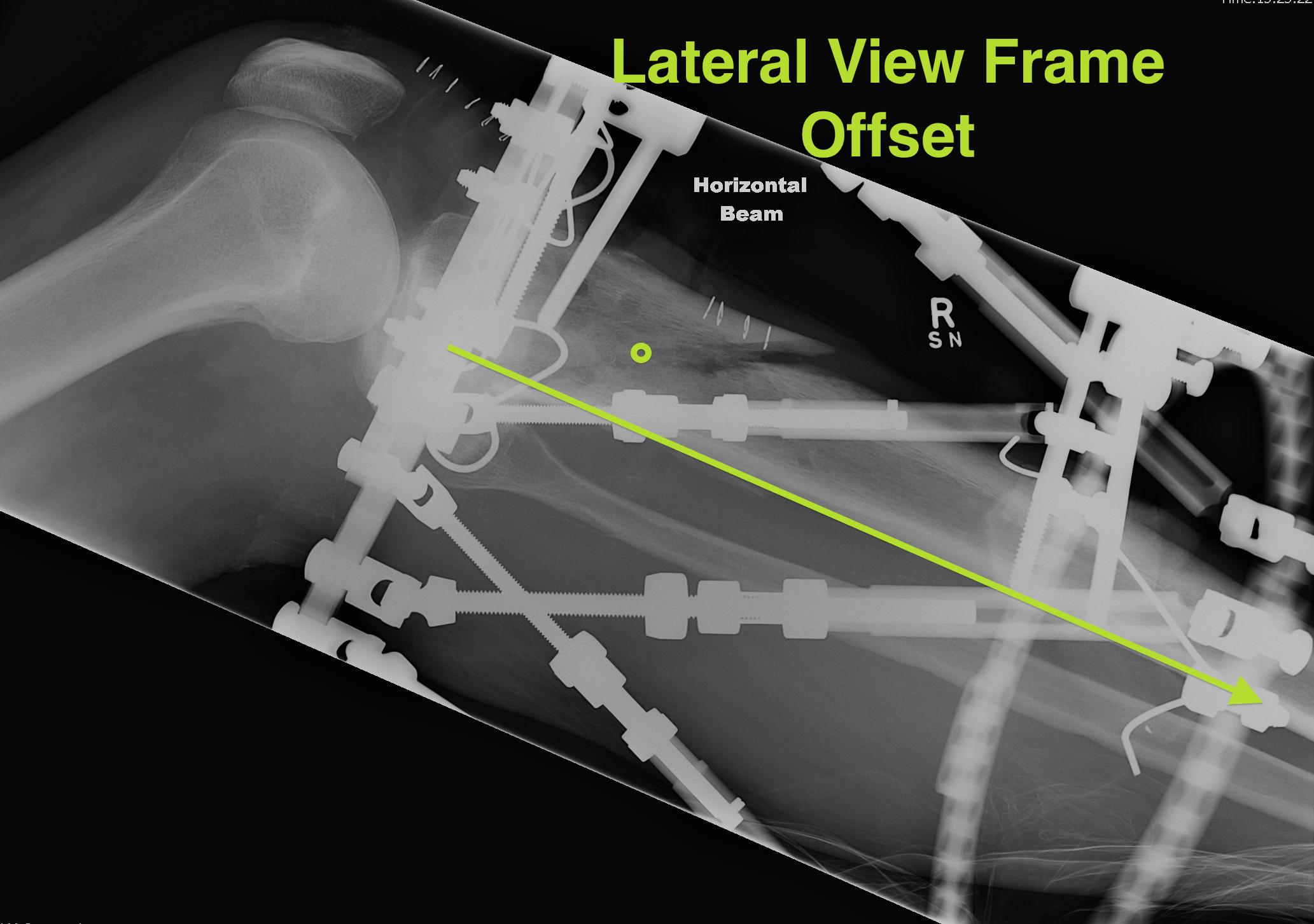
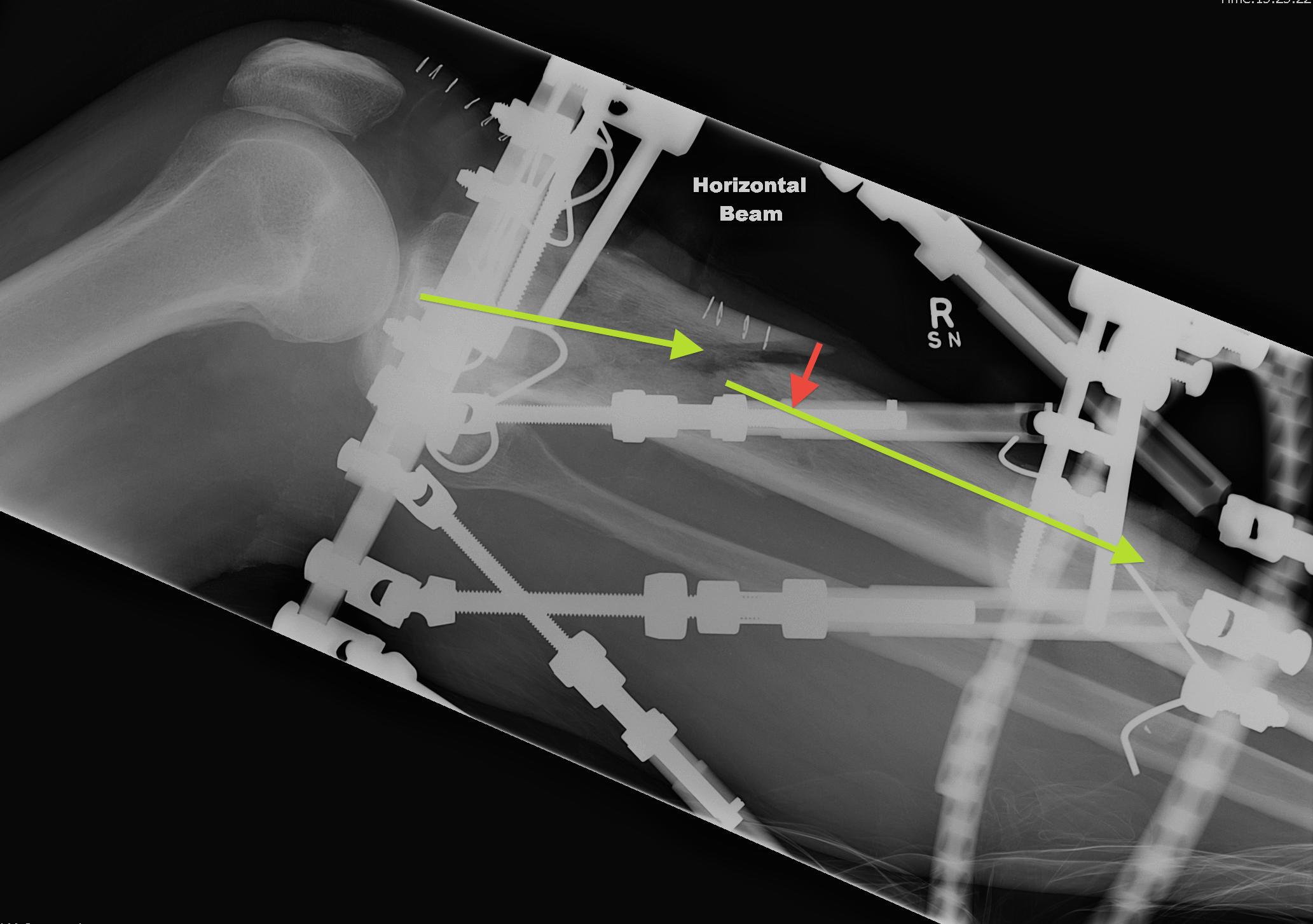
Measure AP and Lateral View Axial Frame Offset
The tibia is not in the centre of the rings
- usually anterior
- need to tell computer where the proximal fragment is in reference to the rings
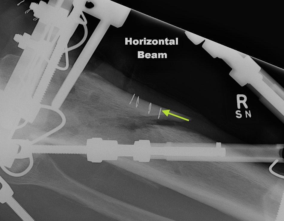
Estimate Rotatory Offset
If Proximal Ring Tab not centred over anterior border of tibia
- need to clinically estimate the difference
- i.e. 5o
Characterise the skeletal deformity
6 measurements required
AP View Angulation + Translation
Lateral View Angulation + Translation
Axial Translation (Shortening)
Axial View Angulation
Clinical
- is the foot externally rotated
- if so, how much
Total Residual Correction
Enter details
- ring size
- strut length
- reference fragment / origin
- frame offset
- 6 measurements
Tell rate of correction
- usually 10 days to 2 weeks
Computer will calculate correction
- assumes correct to neutral
- print daily strut changes requires
Example