Definition
Benign, malformed vascular lesions
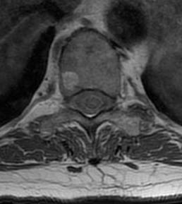
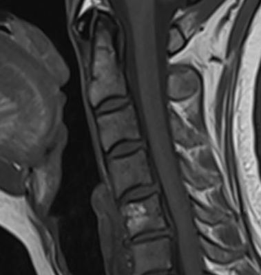
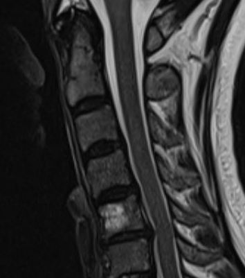
Intraosseous Hemangioma
Epidemiology
Vertebral column
Skull
Long bones rare
Clinical
Usually asymptomatic
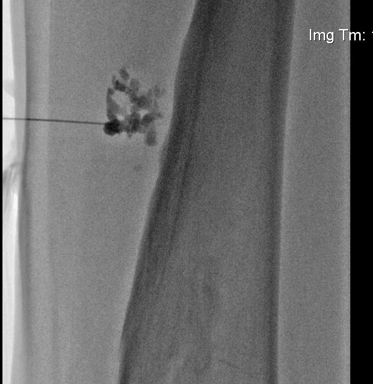
Xray
Characteristic finding is prominent trabecula pattern
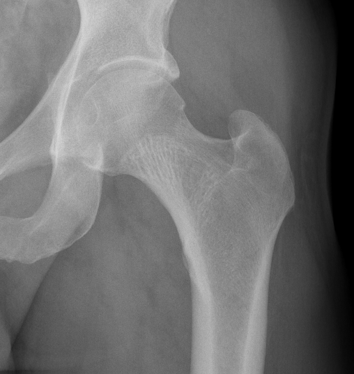
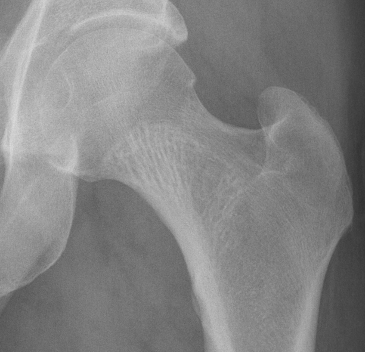
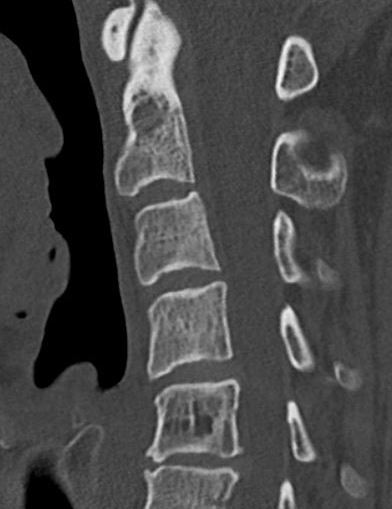
Spine
- characteristic honeycomb appearance
- "jail bar"
CT
Characteristic finding is "polka dots"
MRI
Usually bright on T1 & T2 images because of fat content
Management
Asymptomatic
Typically nonoperative
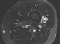
Soft tissue Hemangioma
Associations
1. Klippel Trenaunay Weber Syndrome
- hemihypertrophy with underlying venous malformations
- secondary to increased angiogenesis
- upper limb, lower limb or both affected
- usually unilateral
2. Maffucci Syndrome
3. Sturge-Weber
- rare congenital / not hereditary
- often facial capillary malformation
- more extensive hemangiomata
- development delay / seizures / hemiparesis
- hemiatrophy
Clinical
Ache
Limb heaviness
Lesions in the skin - distended bluish discolouration
Deeper intramuscular lesions present as a tender mass
X-ray
Often small calcified nodules / phleboliths
- circular pattern with a radiolucent center (due to recanalization)
- same as those seen in the pelvic veins of multiparous females
CT
Polka dot appearance due to section through the vessels
These lesions often penetrate the bone and have a large soft tissue component
MRI
Exceedingly bright signal due to the high fluid content of the lesion
Management
Non-operative
- stockings
- simple analgesia
Operative
- embolization
- resection