Definition
Loss of normal ligamentous and / or bony constraints of wrist
Anatomy
Overall alignment maintained by extrinsic and intrinsic ligaments
1. Intrinsic ligaments
Carpal bone to carpal bone
- support the lunate in a balanced position
A. Scapho-lunate ligaments
SL ligament can be divided into three different zones
- dorsal ligamentous zone (structurally the most important)
- palmar ligamentous zone
- proximal membranous fibrocartilaginous zone
B. Luno-triquetral ligaments
- also 3 components
- volar most strong
2. Extrinsic Ligaments
Radius to carpus
- obliquely oriented
- resist the tendency of the carpus to migrate ulnarly and palmarly
A. Palmar extrinsic ligaments
A. Radioscaphocapitate ligament
B. Radiolunate ligament
C. Radioscapholunate ligament
- probably just a vascular fold
D. Ulnocarpal ligaments
E. Lunotriquetral ligament
Space of Poirier
- weak area of the palmar ligaments
B. Dorsal Extrinsics
A. Dorsal radiotriquetral ligament / Dorsal radiocarpal ligament (DRC)
B. Dorsal radioulnar ligament
C. Triquetroscaphoid ligament / Dorsal intercarpal ligament (DIC)
No tendons attach to proximal row
Note:
- acess to dorsal carpus
- raise a radially based flap
- between radiotriquetral and triquetroscaphoid
- between DRC and DIC
Biomechanics
Motion
Capitate is centre of rotation
Flexion / Extension
- 120o
- 50% midcarpal
- 50% radiocarpal
Radial / ulna deviation
- 60% midcarpal
- 40% radiocarpal
Radial deviation
- 20o
- proximal row and scaphoid flexes
Ulnar deviation
- 30o
- proximal row and scaphoid extends
Load transfer
Radius 80%
Ulna 20% (all via TFCC)
Pathology
Division of the scapholunate ligament
- allows the lunate to follow the triquetrum's unrestrained position of extension
- dorsal intercalated segmental instability pattern (DISI)
- scaphoid flexes, lunate extends
Lunotriquetral ligament disruption
- allows the lunate to follow the scaphoid into its position of unrestrained flexion
- lunate flexes
- volar intercalated segmental instability pattern (VlSI)
Classification of Carpal Instabilities (Amadio)
I. Carpal instability dissociative (CID)
Transverse injury
Injury inter-osseous ligaments
- within the carpal rows
- disassociative rather than associative motion between the bones of each row
A.Dorsiflexion (DISI)
- scapholunate ligament injury
B. Palmar flexion (VISI)
- triquetrolunate injury
II. Carpal instability non-dissociative (CIND)
Transverse injury
Normal associative motion between the bones of each carpal row
- the dissociation is between rows
A. Radiocarpal Dislocation
B. Midcarpal
C. Ulnar Translocation
CIND DISI
Secondary to radial malunion
- treat with radial osteotomy if symptomatic
CIND VISI
Secondary to ligamentous laxity
- non operative treatment
- no progression to OA
Whole proximal row is flexed
- lunate triangular
- scaphoid cortical ring sign
- no SL disassociation
III. Carpal instability complex (CIC)
Hyperextension injury
As the hand is forced into hyperextension
- ulnar deviation and intercarpal supination
- the ligamentous disruption
Mayfield Cadaver study
- extend, ulna deviate, supinate
Stage 1
- SL dissociation
Stage 2
- CL dissociation
- capitate dislocates
Stage 3
- LT dissociation
Stage 4
- Lunate dislocates
Types
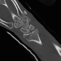
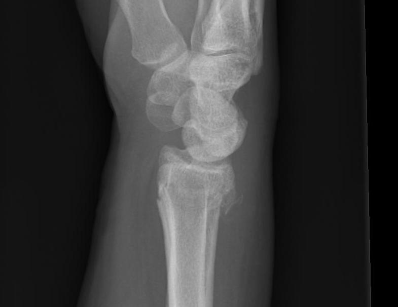
A. Perilunate Dislocation
1. Dorsal (10%)
2. Volar (90%)
B. Trans-scaphoid Perilunate
IV. Carpal instability longitudinal (axial)
Longitudinal injury
The carpus may also be disrupted in a longitudinal fashion, as opposed to the perilunate transverse pattern
Classification
A. Axial Ulnar (AU)
B. Axial Radial (AR)
C. Axial Ulnar-Radial (AUR) / Combined
These are severe injuries
- crush, blast or compression
- may be open injuries
- not a diagnostic dilemma
Usually wrist is split into two columns
- metacarpals follow their corresponding carpus
Management
Deal with wounds and nerve / tendon injuries
CTD
K wire fixation
Greater and Lesser Arc Injuries
Greater arc injury
- fracture-dislocation of the scaphoid, capitate, hamate, triquetrum
- may include radial styloid
Lesser arc injury
- a pure ligamentous injury
- around the lunate