Definition
Volar Intercalated Segmental Instability
- secondary to injury to the lunate-triquetral ligament
Epidemiology
Less common
Aetiology
Caused by fall on outstretched extended wrist
- hypothenar eminence strikes ground first
- isolated LT ligament injury
Can be part of perilunate dislocation
- SL heals
- residual LT laxity
Anatomy
LT ligament
- also C shaped
- strongest palmar
Pathomechanics
Normally
- scaphoid imparts a flexion moment on proximal row
- triquetrum imparts an extension moment
- balanced by ligamentous attachments to lunate
Palmarflexion of lunate with dorsiflexion of triquetrum
Probably need injury to dorsal extrinsics to impart static collapse
- DRC ligament (radio-triquetral)
- ulnocarpal ligament
Classification
CID
Static
Dynamic
CIND VISI
Secondary to ligamentous laxity
- seen in teenage girls
- clunk on radial and ulna deviation with axial compression
Whole proximal row is flexed
- lunate triangular
- scaphoid cortical ring sign
- no SL disassociation
Non operative treatment
- no progression to OA
Symptoms
History of injury
Pain on ulnar side of wrist
Weakness of wrist
Signs
Swelling and tenderness over triquetro-lunate joint
Ulna deviation / pronation / axial compression
- pain and clicks
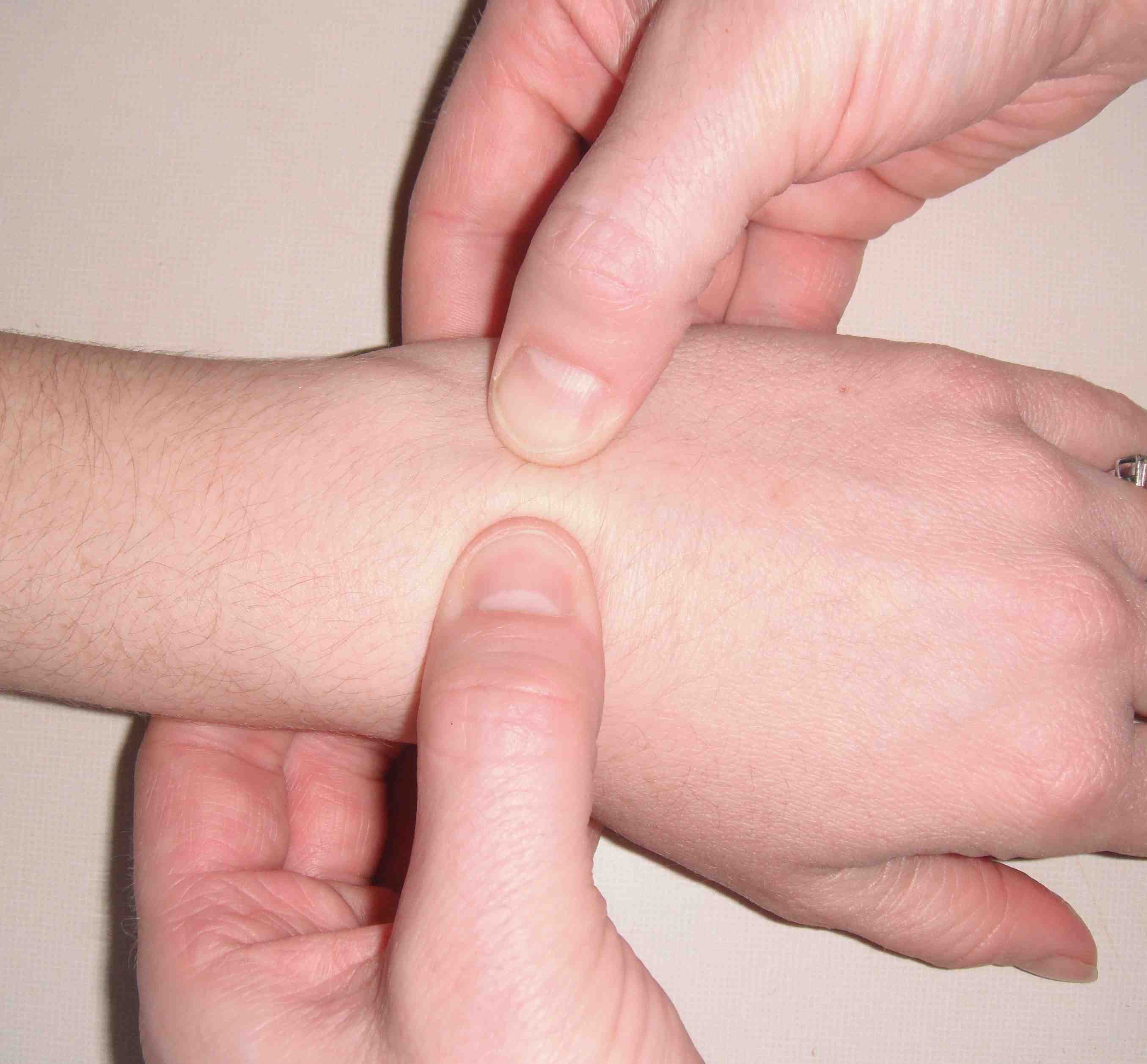
Reagan Ballotment
- Triquetro-lunate ballottement
- pisiform-triquetral with thumb and index finger
- lunate with other hand
DDx
DRUJ instability
TFCC tear
Ulna head OA
Pisiform triquetral OA
Hamate fracture
ECU subluxation
AP Xray
Palmarflexion of scaphoid
- Scaphoid shortened
- Ring sign
Palmarflexion of lunate
- Appears triangular
- Triquetrum distally displaced
Broken Shenton's line (of proximal carpal row)
Lateral Xray
Decreased scapholunate angle
- < 30o
Palmarflexion of lunate
- capitate - lunate angle > 10o
- radio - lunate angle > 10o
Arthroscopy
Diagnostic and therapeutic
Management
Early
Options
A. Repair
- dorsal approach
- restore LT orientation with K wires
- repair ligament with intra-osseous sutures
B Reconstruct with ECU
- if insufficient ligament for repair
- radial half of ECU
- pass through drill holes
Late
> 6 weeks
Lunate-triquetral fusion
- very difficult
- high failure with k wires
- need compression screws
- insert bone graft