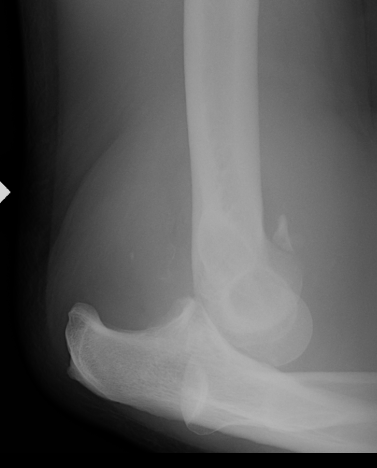
Acute Elbow Dislocation Management
1. Reduction under IV / conscious sedation
- assistant applies traction in slight flexion
- second person corrects lateral displacement by manipulating olecranon medially
- flexion to 90o
2. Post reduction assess stability
- stable if can extend to within 30 - 40o without instability
- if unstable, pronate forearm and see if can extend to within 30 - 40o (MCL intact)
- if unstable pronated with elbow < 45o extended, elbow will need surgery
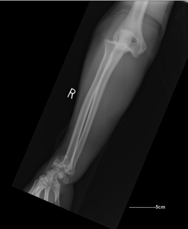
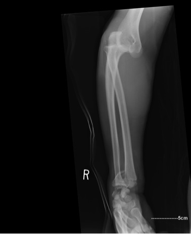
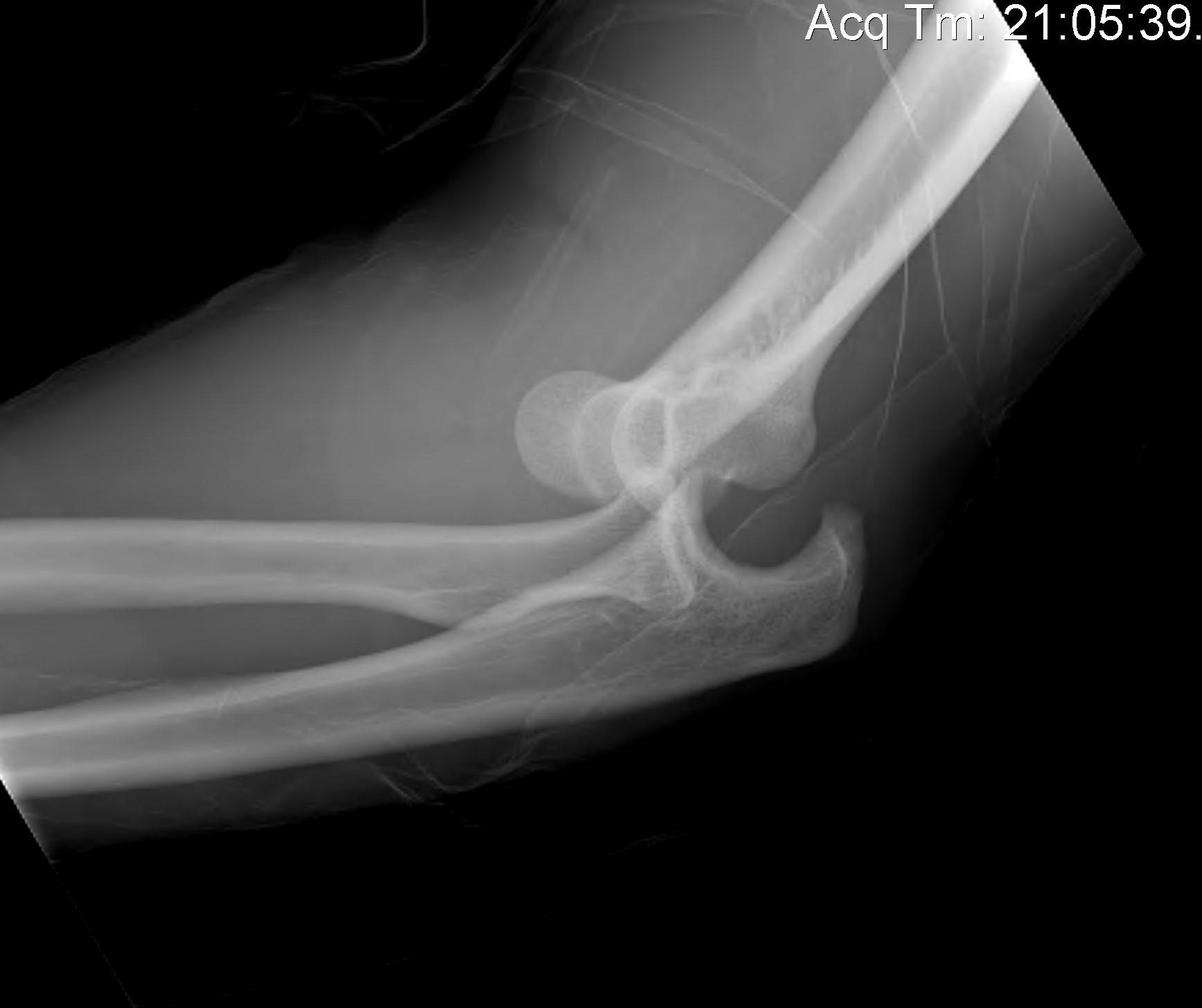
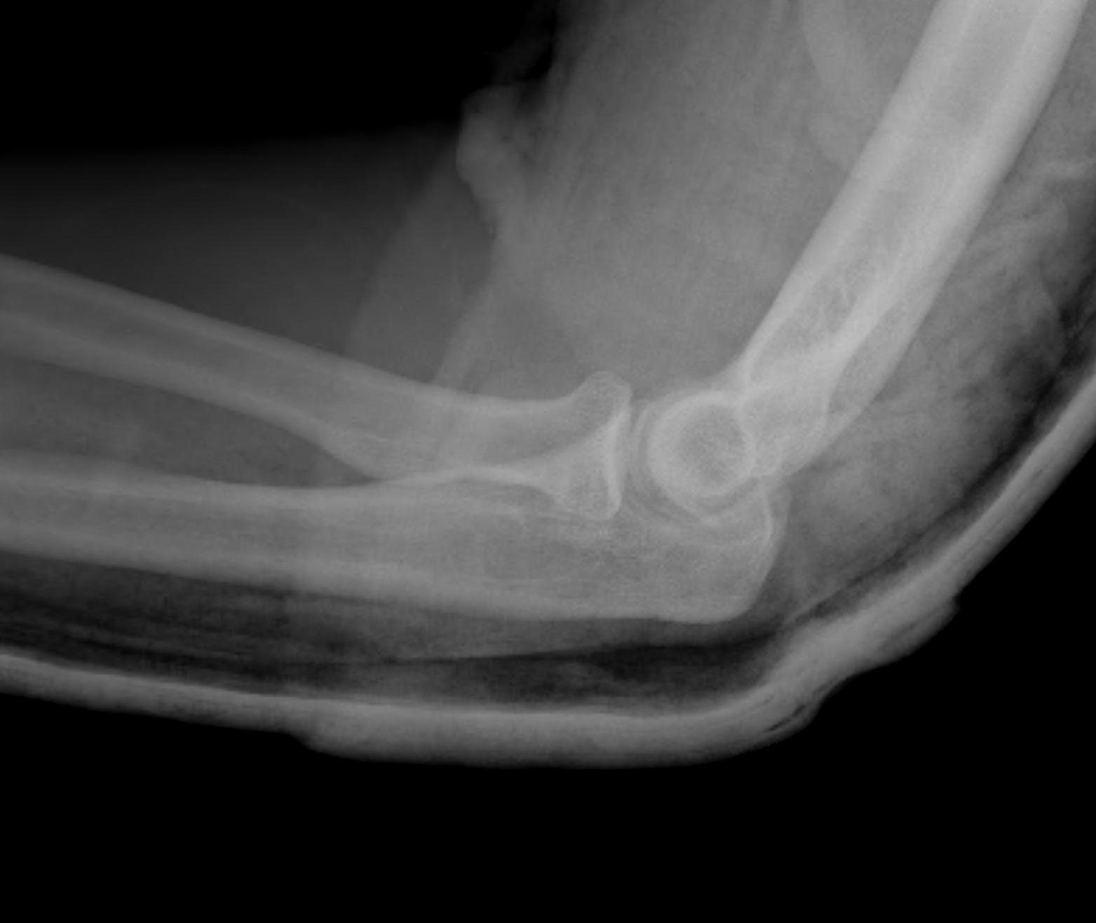
3. Confirm concentric reduction
- 2 view check x-rays mandatory
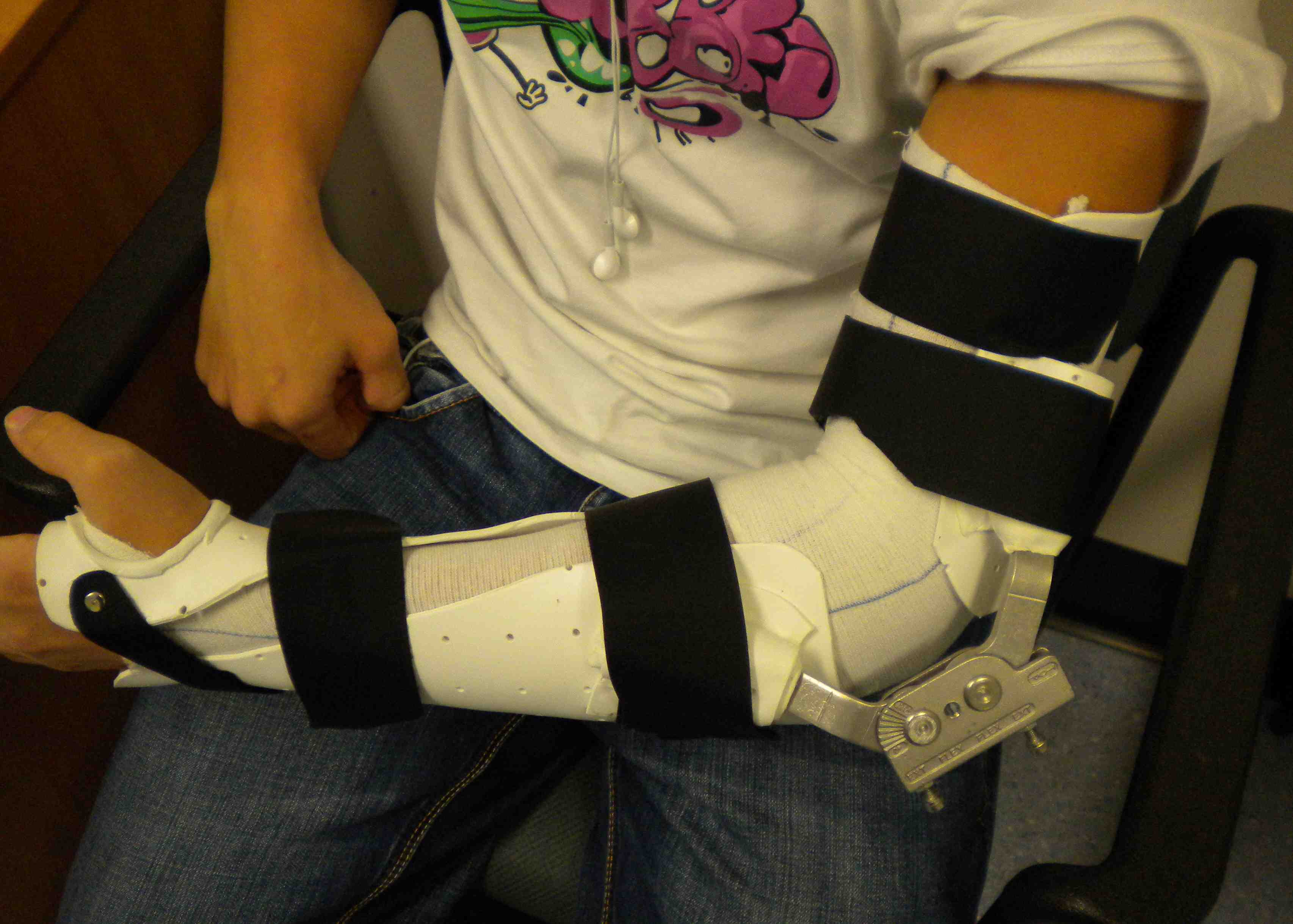
4. Stable elbow
- manage in POP 90o 2 weeks
- weekly check xray
- then begin ROM exercises
Management Problems
A. Simple Elbow Dislocation
B. Complex Elbow Dislocation
- radial head fracture
- coronoid process fracture
- Terrible Triad (MCL / coronoid / radial head)
- olecranon fracture +/- radial head +/- coronoid
- capitellar fractures
Note
- difficult problem
- need to prepared at all times to
- ORIF / replace radial head
- repair / reconstruct LCL
- ORIF / suture coronoid
- repair MCL
- apply external fixator
1. Simple Elbow Dislocation
A. Stable Simple Elbow Dislocation
Management
Reduce
Assess Stability
- OT if unstable > 45o in pronation
X-ray weekly
Mobilise 2 - 3 weeks
If FFD at 6/52 > 40o
- night extension splint
- turnbuckle elbow extension splints
Josefsson et al 1987 JBJS AM
- randomised 30 patients with elbow dislocations
- non-operative group 2 weeks in plaster at 90°
- operative group had ruptures of both collaterals / most had avulsions from the humeral epicondyles
- no difference in outcome between the two groups regardless of initial stability
- loss of extension was commonest complication
- seen 50% more in operative group
B. Unstable simple elbow dislocation
Uncommon but not rare
- may be intact medially
- avulsed LCL and CEO
Algorithm
1. Kocher approach & Reconstruct / Repair LCL + CEO
- lateral ulna collateral ligament is usually avulsed from lateral condyle
- centre of rotation is centre of capitellum
- place suture anchor
- repair anconeus and ECU over top
- +/- reconstruct / augment with slip Palmaris if required
- ROM brace
2. Elbow still unstable / address MCL
- usually avulsed from medial epicondyle
- usually can do direct repair / suture anchors
- mid-substance probably have to reconstruct with Palmaris
Medial approach centred on medial epicondyle
- locate, mobilise and protect ulna nerve
- proximally between brachialis and triceps
- distally between pronator teres and brachialis
- can reflect PT
- protect median nerve distally
C. Chronic Simple Elbow dislocation
Missed injury / delayed presentation
- open reduction
- removal scar tissue
- repair / reconstruction LCL
- +/- hinged external fixation
2. Dislocation with Radial Head Fracture
Manage as per radial head classification
Hotchkiss Modified Mason class (R&G)
Type I
Non / minimally (<2mm) displaced fracture of head
- forearm rotation (pronation/supination) is limited only by acute pain and swelling
- diagnose by LA injection and full pronation and supination
Non operative treatment
Type II
Displaced fracture of the head or neck
- > 2mm and amenable to fixation
Motion may be mechanically limited with or without significant joint incongruity
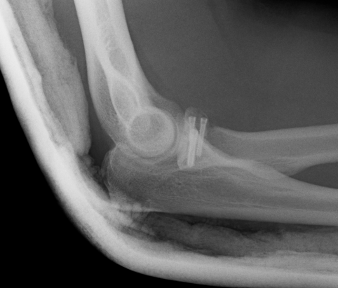
Management
- Kocher approach
- ORIF
- LCL repair / reconstruction
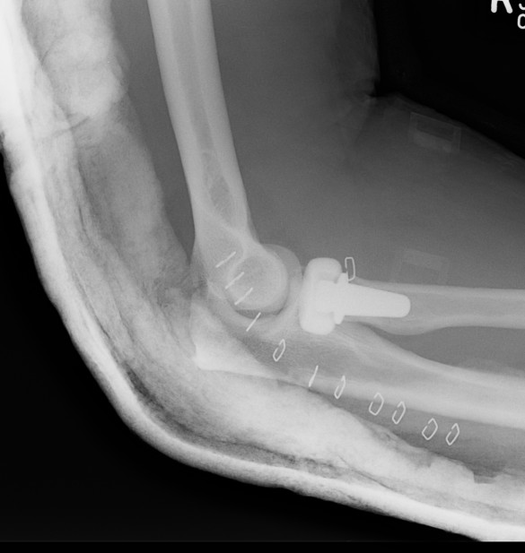
Type III
Severely comminuted fracture of the radial head and neck
- not reconstructable
- Titanium replacement
Ashwood et al JBJS Am 2004
- 16 patients titanium monoblock radial head
- 81% G/E at 2 years
Radial Neck Fracture
Morrey et al J Orthop Trauma
- concern regarding loss of rotation with plating
- prefer to ORIF with oblique screws or radial head replacement
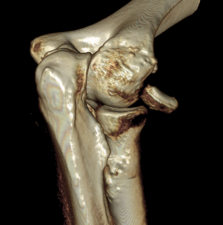
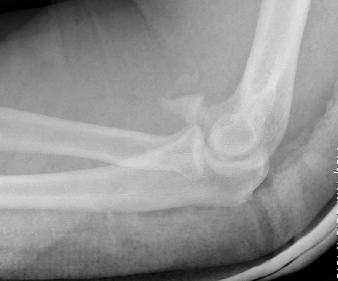
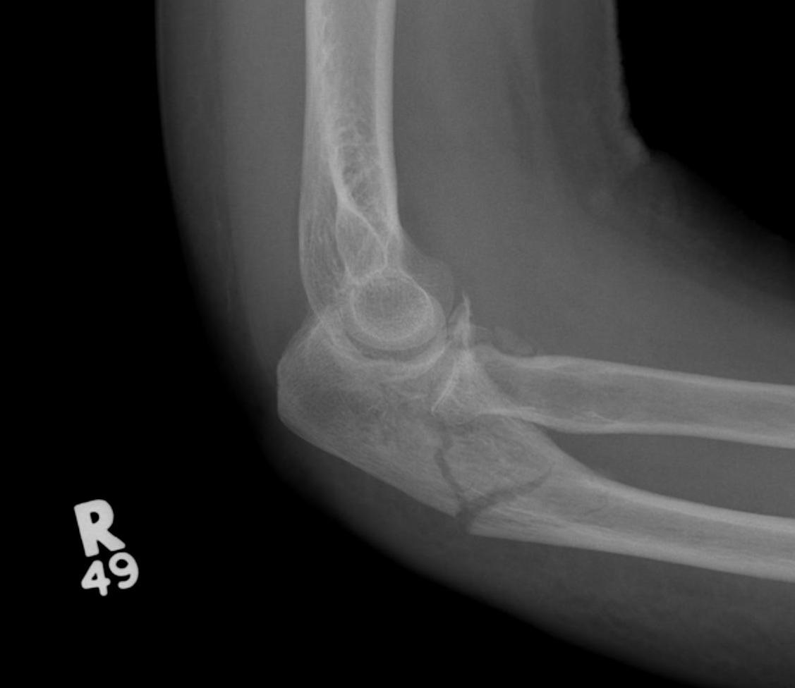
3. Dislocation with Coronoid Fracture
The coronoid is the most important portion of ulno-humeral articulation
Reasons
- provides anterior buttress
- attachment of capsule and brachialis
- anterior band of the MCL attaches to it
Manage as per Regan and Morrey Classification
- ORIF / repair type I / II
Regan and Morrey Classification
Type I
- stable as nothing attaches to tip
- shear fracture, not avulsion fracture
Type II
- 50% coronoid
- elbow usually unstable / ORIF or suture
Type III
- > 50%
- uncommon
- can be comminuted
- ORIF or suture
Approach
Universal posterior approach
- single posterior skin incision
- elevate flaps laterally and medially as required
- lateral approach to repair ulna LCL
- medial approach to repair coronoid
Medial approach
- isolate and protect ulna nerve
- elevation of ulna origin of flexor pronator group anterior to FCU
- important if fracture is medial
Fixation
1. Screw / buttress plate
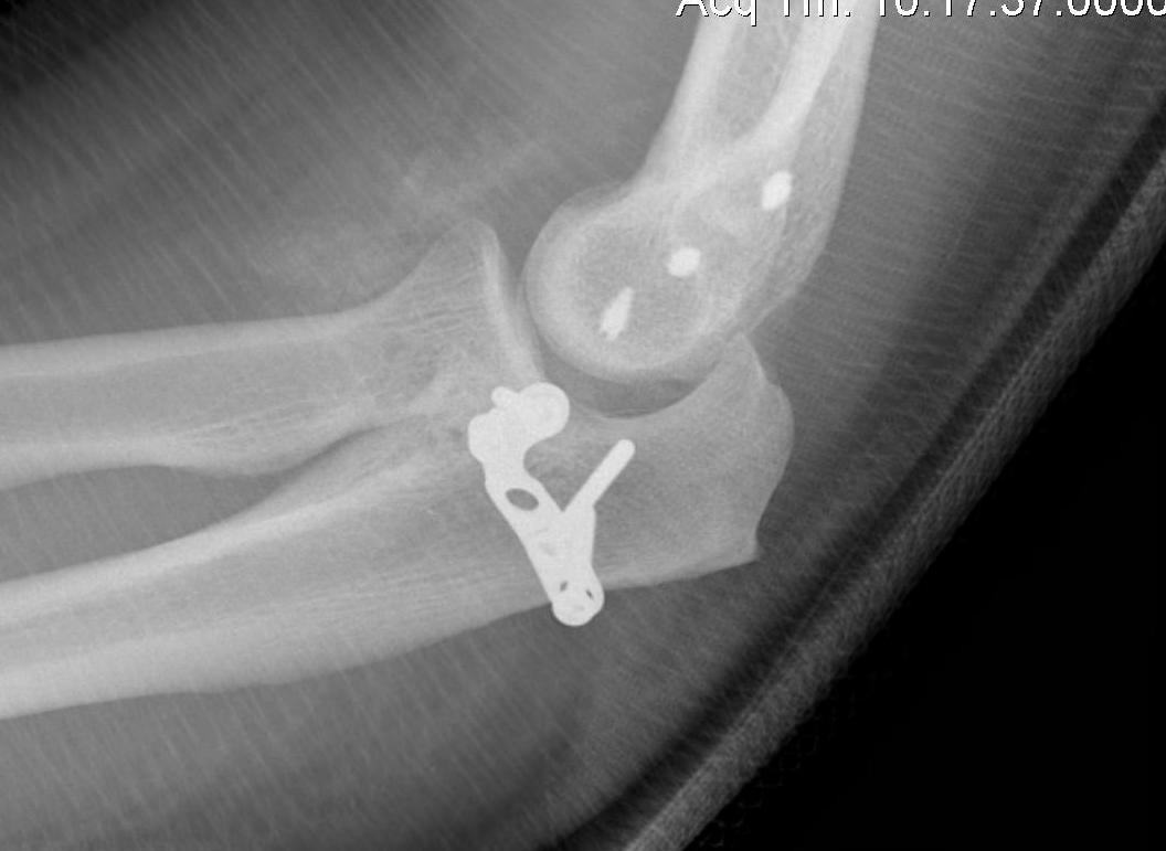
2. Sutures through capsule / Lasso repair
- tie over drill holes through olecranon / endobutton
3. Reconstruct with radial head, iliac crest, or allograft
Note: Acknowledged by world class names as being difficult
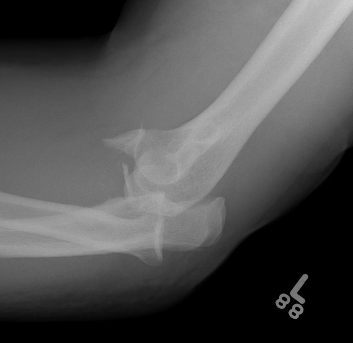
4. Dislocation + Terrible Triad
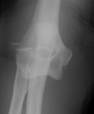
Definition
- radial head fracture + coronoid fracture + MCL
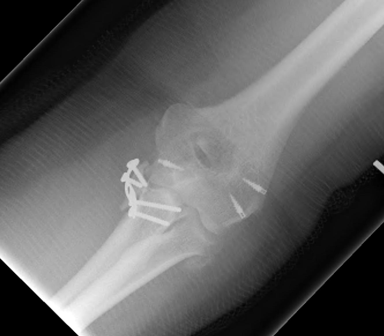
Surgical Algorigthm
Universal Posterior Approach
1. Type 2 radial head
- Kocher approach
- ORIF
- repair / reconstruct ulna LCL
- reassess stability
- if unstable, additional medial approach
- isolate and protect ulna nerve
- if type II / III coronoid elevate CFO and ORIF / suture
- repair / reconstruct MCL
- assess stability
- rarely may require external fixator
2. Type 3 radial head
- Kocher approach
- excise radial head
- attempt ORIF / suture coronoid process through this gap
- unless large anteromedial fracture which is best treated with anteromedial buttress plate
- replace radial head
- repair / reconstruct LCL
- reassess stability
- may then need medial approach and MCL repair / reconstruction
- reassess stability
- may need hinged external fixator
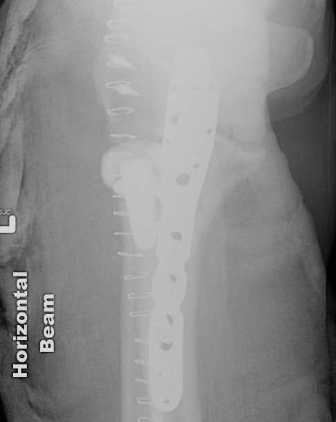
5. Dislocation with Olecranon Fracture +/- Coronoid Fracture +/- Radial Head Fracture
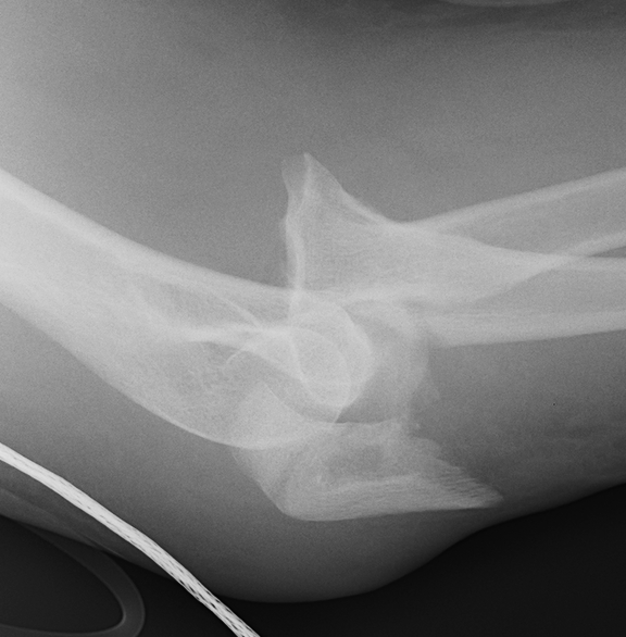
A. Anterior / Trans Olecranon Fracture Dislocations
Less common, better outcomes because
- coronoid fragment usually larger / easier to ORIF
- collaterals often intact
- radial head often intact
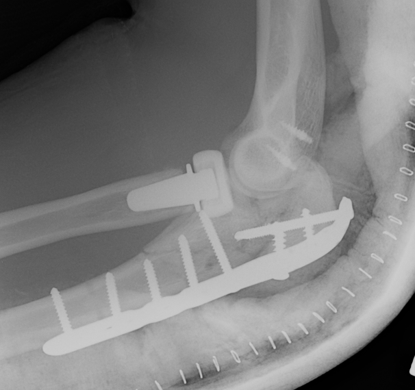
Management
- universal posterior approach
- ORIF / suture coronoid through olecranon fracture
- TBW or plate for olecranon fracture
- can repair coronoid with lag screw from olecranon plate
- Kocher approach
- ORIF / replace radial head
- repair / reconstruct LCL
- reassess stability
- +/- repair reconstruct MCL
B. Posterior Monteggia Fracture
More common, worse outcome because
- LCL more likely to be ruptured as well
- coronoid more likely to be comminuted
- radial head fracture
Management
- ORIF coronoid through olecranon fracture
- ORIF olecranon (often plate as distal to centre of rotation of elbow)
- +/- ORIF /replace radial head
- +/- repair / reconstruct LCL
- +/- hinged fixator
6. Other
Dislocation with distal radius fracture